We apologize for Proteopedia being slow to respond. For the past two years, a new implementation of Proteopedia has been being built. Soon, it will replace this 18-year old system. All existing content will be moved to the new system at a date that will be announced here.
ExbD
From Proteopedia
(Difference between revisions)
| Line 1: | Line 1: | ||
| + | <StructureSection load='2pfu' size='350' side='right' scene='' caption=''> | ||
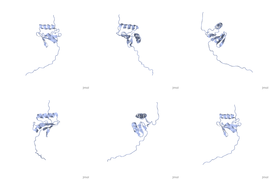
[[Image:ExbD.jpg|300px|left|thumb| The Structure of ExbD<ref name='Kampfenkel'>PMID: 1644779</ref>]] | [[Image:ExbD.jpg|300px|left|thumb| The Structure of ExbD<ref name='Kampfenkel'>PMID: 1644779</ref>]] | ||
==Structure== | ==Structure== | ||
| - | + | ||
'''ExbD''' has a single transmembrane domain, with residues 1 to 22 on the cytoplasmic side and 44 to 141 in the periplasm (''see'' 3D structure 2PFU). Residues 23 to 43 are within the cytoplasmic membrane and it is in this region, from residues 18 to 43, that the only hydrophobic residues in ExbD can be found<ref name='Kampfenkel'>PMID: 1644779</ref>. | '''ExbD''' has a single transmembrane domain, with residues 1 to 22 on the cytoplasmic side and 44 to 141 in the periplasm (''see'' 3D structure 2PFU). Residues 23 to 43 are within the cytoplasmic membrane and it is in this region, from residues 18 to 43, that the only hydrophobic residues in ExbD can be found<ref name='Kampfenkel'>PMID: 1644779</ref>. | ||
| Line 12: | Line 13: | ||
The activity of ExbD can be affected with mutations of the single charged amino acid (here D25N) which lies close to the transmembrane region. This can also be said of the other transmembrane proteins ExbB, TolQ and TolR. | The activity of ExbD can be affected with mutations of the single charged amino acid (here D25N) which lies close to the transmembrane region. This can also be said of the other transmembrane proteins ExbB, TolQ and TolR. | ||
| - | + | </StructureSection> | |
| - | ==3D structures of | + | ==3D structures of ExbD== |
[[2pfu]] – EXBD periplasmic domain – ''Escherichia coli'' – NMR | [[2pfu]] – EXBD periplasmic domain – ''Escherichia coli'' – NMR | ||
Revision as of 08:19, 6 June 2016
| |||||||||||
3D structures of ExbD
2pfu – EXBD periplasmic domain – Escherichia coli – NMR
References
- ↑ 1.0 1.1 1.2 Kampfenkel K, Braun V. Membrane topology of the Escherichia coli ExbD protein. J Bacteriol. 1992 Aug;174(16):5485-7. PMID:1644779
- ↑ Held KG, Postle K. ExbB and ExbD do not function independently in TonB-dependent energy transduction. J Bacteriol. 2002 Sep;184(18):5170-3. PMID:12193634
- ↑ Braun V, Herrmann C. Point mutations in transmembrane helices 2 and 3 of ExbB and TolQ affect their activities in Escherichia coli K-12. J Bacteriol. 2004 Jul;186(13):4402-6. PMID:15205446 doi:10.1128/JB.186.13.4402-4406.2004


