User:R. Jeremy Johnson/Glucagon Receptor
From Proteopedia
(Difference between revisions)
| Line 3: | Line 3: | ||
==Class B GPCRs== | ==Class B GPCRs== | ||
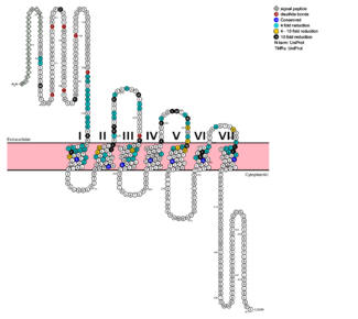
| - | G protein coupled receptors (GPCRs) are the largest class of integral membrane proteins. GPCRs are divided into five families; the rhodopsin family (class A), the secretin family (class B), the glutamate family (class C), the frizzled/taste family (class F), and the adhesion family.<ref name= "Zhang 2006"/> Roughly 5% of the human genome encodes g protein-coupled receptors, which are responsible for the transduction of endogenous signals and the instigation of cellular responses.<ref name= "Zhang 2006"/> All GPCRs contain a similar seven α-helical transmembrane domain <scene name='72/727091/Full_Structure_with_Labels/1'>(TMD or 7TMD)</scene> that once bound to its ligand, undergoes a conformational change and tranduces a signal to coupled, heterotrimeric G proteins. The initiation of intracellular signal pathways occur in response to stimuli such as light, | + | G protein coupled receptors (GPCRs) are the largest class of integral membrane proteins. GPCRs are divided into five families; the rhodopsin family (class A), the secretin family (class B), the glutamate family (class C), the frizzled/taste family (class F), and the adhesion family.<ref name= "Zhang 2006"/> Roughly 5% of the human genome encodes g protein-coupled receptors, which are responsible for the transduction of endogenous signals and the instigation of cellular responses.<ref name= "Zhang 2006"/> All GPCRs contain a similar seven α-helical transmembrane domain <scene name='72/727091/Full_Structure_with_Labels/1'>(TMD or 7TMD)</scene> that once bound to its ligand, undergoes a conformational change and tranduces a signal to coupled, heterotrimeric G proteins. The initiation of intracellular signal pathways occur in response to stimuli such as light, Ca<sup>2+</sup>, amino acids, nucleotides, odorants, peptides, and other proteins, [https://en.wikipedia.org/wiki/G_protein%E2%80%93coupled_receptor#Physiological_roles and accomplishes many interesting physiological roles]. <ref name= "Zhang 2006">DOI 10.1371/journal.pcbi.0020013</ref> The '''human glucagon receptor''' ('''GCGR''') is one of 15 secretin-like, or Class B, [https://en.wikipedia.org/wiki/G_protein%E2%80%93coupled_receptor G-protein coupled receptors] (GPCRs). Like other GPCRs, it has a <scene name='72/721538/7tm_labeled_helicies/3'>7 trans-membrane </scene> helical domain and a globular N-terminus <scene name='72/721538/Ecd/2'>extracellular domain</scene> (ECD). [[Image:Protter GLR HUMAN.png |325 px|left|thumb|'''Figure 1''': Snake Plot of GCGR TMD. Residues of particular importance in glucagon binding affinity are found in green, yellow, and black. Residues in red are the location of critical disulfide bonds, while blue residues were found to be highly conserved across all class B GPCRs.<ref name= "Siu 2013"/>]] |
| - | Class B GPCRs contain 15 distinct receptors for peptide hormones and generate their signal pathway through the activation of adenylate cyclase (AC) which increases the intracellular concentration of cAMP, inositol phosphate, and calcium levels. <ref>DOI 10.1111/bph.12689</ref> These secondary messengers are essential elements of intracellular signal cascades for human diseases including type II diabetes mellitus, osteoporosis, obesity, cancer, neurological degeneration, cardiovascular diseases, headaches, and psychiatric disorders; making their regulation through drug targeting of particular interest as disease targets. <ref name= "Hollenstein 2014">DOI 10.1016/j.tips.2013.11.001</ref> Structural approaches to the development of agonists and antagonists have however been hampered by the lack of accurate Class B TMD visualizations. Recent crystal | + | Class B GPCRs contain 15 distinct receptors for peptide hormones and generate their signal pathway through the activation of adenylate cyclase (AC) which increases the intracellular concentration of cAMP, inositol phosphate, and calcium levels. <ref>DOI 10.1111/bph.12689</ref> These secondary messengers are essential elements of intracellular signal cascades for human diseases including type II diabetes mellitus, osteoporosis, obesity, cancer, neurological degeneration, cardiovascular diseases, headaches, and psychiatric disorders; making their regulation through drug targeting of particular interest as disease targets. <ref name= "Hollenstein 2014">DOI 10.1016/j.tips.2013.11.001</ref> Structural approaches to the development of agonists and antagonists have however been hampered by the lack of accurate Class B TMD visualizations. Recent crystal structures of corticoptropin-releasing factor receptor 1 (PDB: 4K5Y) and human glucagon receptor (PDB: 4L6R) provide a comparison to more well-studied class A GPCRs. <ref name= "Hollenstein 2013">DOI 10.1038/nature12357</ref><ref name= "Siu 2013">DOI 10.1038/nature12393</ref> |
==Structures of Class A vs. Class B GPCRs== | ==Structures of Class A vs. Class B GPCRs== | ||
| - | Class A | + | Class A and class B glucagon receptors share less than fifteen percent sequence homology, but both share a 7TM domain.<ref name= "Hollenstein 2013"/> Comparison of the <scene name='72/721536/Final_1st_image/1'>class B 7TM</scene> helices to that of the <scene name='72/721536/Final_class_a_7tm/1'>class A 7TM</scene> helices showed that the general orientation and positioning of the [https://en.wikipedia.org/wiki/Alpha_helix alpha helices] are conserved through both classes. Detailed structural alignments of the two GPCR subclasses revealed deviations in the transmembrane region.<ref name= "Siu 2013"/> For instance, the N-terminal end of <scene name='72/721536/Final_helix_1/2'>helix one</scene> in class B GCGR is longer than any known class A GPCR structure and stretches three supplementary helical turns above the extracellular (EC) membrane boundary. This region is referred to as the <scene name='72/721535/stalk/1'>stalk</scene>. The stalk is involved in glucagon binding and helps in defining the orientation of the ECD with respect to the 7TM domain.<ref name= "Siu 2013"/> Also specific to class B GPCRs, a [https://en.wikipedia.org/wiki/Glycine Gly] residue at position 393 induces a <scene name='72/721535/Helical_bend/4'>bend in helix VII</scene>; this bend is stabilized by the [http://chemwiki.ucdavis.edu/Core/Physical_Chemistry/Physical_Properties_of_Matter/Atomic_and_Molecular_Properties/Intermolecular_Forces/Hydrophobic_Interactions hydrophobic interaction] between the <scene name='72/721535/Gly_393_phe_184/2'> glycine 393 and phenylalanine 184</scene>. One of the most distinguishable characteristics of the class B 7TM is the <scene name='72/721536/Class_b_helix_8_tilt_finals/1'>helix VIII tilt</scene> of 25 degrees and its length compared to that of <scene name='72/721536/Class_a_helix_vii_tilt/2'>class A helix VIII tilt</scene>, which is much shorter. This helical tilt results from [https://en.wikipedia.org/wiki/Phenylalanine Glu] 406 in helix VIII that is fully conserved in secretin-like receptors and forms two interhelical <scene name='72/721537/Salt_bridges/2'>salt bridges</scene> with [https://simple.wikipedia.org/wiki/Conserved_sequence conserved residues] [https://en.wikipedia.org/wiki/Arginine Arg] 173 and Arg 346.<ref name= "Siu 2013"/> Despite these differences, a vital region that is conserved in both class B and class A receptors is the [https://en.wikipedia.org/wiki/Disulfide disulfide bond] between <scene name='72/721535/Disulfide_bond_notspin_actual/2'>Cys 294 and Cys 224</scene> in extracellular loop two (ECL2). This bond stabilizes the receptors entire 7TM fold. Lastly, the locations of the extracellular tips for class B glucagon receptors allow for a much wider and deeper [https://en.wikipedia.org/wiki/Ligand_(biochemistry) ligand-binding pocket] than any of the class A GPCRs.<ref name= "Siu 2013"/> While interfacial interactions are not unique to class B receptors, part of the interface <scene name='72/721537/Helical_interactions_vi-v-iii/2'>stabilization</scene> between helices VI, V, and III includes a Class B-specific [https://en.wikipedia.org/wiki/Hydrogen_bond hydrogen bond] between [https://en.wikipedia.org/wiki/Asparagine Asn] 318 of Helix V and [https://en.wikipedia.org/wiki/Leucine Leu] 242 of Helix III.<ref name= "Siu 2013"/> |
===How These Structures Lead to Function=== | ===How These Structures Lead to Function=== | ||
| Line 17: | Line 17: | ||
The [http://sbkb.org/fs/glucagon-receptor glucagon class B GPCR (GCGR)] is involved in glucose homeostasis through the binding of the signal peptide glucagon. Glucagon is released from pancreatic α-cells when blood glucose levels fall after a period of fasting or several hours following intake of dietary carbohydrates.<ref name = 'Lehninger'/> Once the peptide hormone is released, it binds to GCGR, a 485 amino acid protein found in the liver, kidney, intestinal smooth muscle, brain, and adipose tissues. <ref name= "Yang 2015">DOI 10.1038/aps.2015.78</ref> Upon binding, signaling is initiated to heterotrimeric G-proteins containing Gαs. <ref name= "Ahren 2009">DOI 10.1038/nrd2782</ref> GCGR can regulate additional signal pathways, including G-proteins of the Gαi family through the adoption of differing receptor conformations. <ref name= "Xu 2009">DOI 10.3109/10799890903295150</ref> | The [http://sbkb.org/fs/glucagon-receptor glucagon class B GPCR (GCGR)] is involved in glucose homeostasis through the binding of the signal peptide glucagon. Glucagon is released from pancreatic α-cells when blood glucose levels fall after a period of fasting or several hours following intake of dietary carbohydrates.<ref name = 'Lehninger'/> Once the peptide hormone is released, it binds to GCGR, a 485 amino acid protein found in the liver, kidney, intestinal smooth muscle, brain, and adipose tissues. <ref name= "Yang 2015">DOI 10.1038/aps.2015.78</ref> Upon binding, signaling is initiated to heterotrimeric G-proteins containing Gαs. <ref name= "Ahren 2009">DOI 10.1038/nrd2782</ref> GCGR can regulate additional signal pathways, including G-proteins of the Gαi family through the adoption of differing receptor conformations. <ref name= "Xu 2009">DOI 10.3109/10799890903295150</ref> | ||
| - | Glucagon's main role is the regulation of blood glucose levels.<ref name = 'Lehninger'/> Glucagon lowers the concentration of fructose 2,6-bisphosphate which is an allosteric inhibitor of the gluconeogenic enzyme fructose 1,6-bisphosphotase and activates phosphofructose kinase 1, which increases glucose levels via glycolysis.<ref name = 'Lehninger'/> Glucagon is also a regulator of the production of cholesterol, which is an energetically intensive process. When energy resources are low, downregulation of cholesterol production begins with glucagon binding to GCGR, which stimulates the phosphorylation of HMG-CoA.<ref name = 'Lehninger'/> HMG-CoA is inactivated by phosphorylation and moderates cholesterol production to conserve energy.<ref name = 'Lehninger'/> Glucagon also takes part in fatty acid mobilization by affecting levels of adipose tissue in the organism. Activation of GCGR by glucagon initiates triacylglycerol breakdown and the phosphorylation of perilipin and lipases via cAMP signal pathways.<ref name = 'Lehninger'/> This allows the body to export fatty acids to the liver and other crucial tissues for energy use and makes more glucose available for use in brain functioning.<ref name = 'Lehninger'>'Lehninger A., Nelson D.N, & Cox M.M. (2008) Lehninger Principles of Biochemistry. W. H. Freeman, fifth edition.' </ref> | + | Glucagon's main role is the regulation of blood glucose levels.<ref name = 'Lehninger'/> Glucagon lowers the concentration of fructose 2,6-bisphosphate, which is an allosteric inhibitor of the gluconeogenic enzyme fructose 1,6-bisphosphotase and activates phosphofructose kinase 1, which increases glucose levels via glycolysis.<ref name = 'Lehninger'/> Glucagon is also a regulator of the production of cholesterol, which is an energetically intensive process. When energy resources are low, downregulation of cholesterol production begins with glucagon binding to GCGR, which stimulates the phosphorylation of HMG-CoA.<ref name = 'Lehninger'/> HMG-CoA is inactivated by phosphorylation and moderates cholesterol production to conserve energy.<ref name = 'Lehninger'/> Glucagon also takes part in fatty acid mobilization by affecting levels of adipose tissue in the organism. Activation of GCGR by glucagon initiates triacylglycerol breakdown and the phosphorylation of perilipin and lipases via cAMP signal pathways.<ref name = 'Lehninger'/> This allows the body to export fatty acids to the liver and other crucial tissues for energy use and makes more glucose available for use in brain functioning.<ref name = 'Lehninger'>'Lehninger A., Nelson D.N, & Cox M.M. (2008) Lehninger Principles of Biochemistry. W. H. Freeman, fifth edition.' </ref> |
=== GCGR-Specific Traits === | === GCGR-Specific Traits === | ||
==== Helix I Stalk Region ==== | ==== Helix I Stalk Region ==== | ||
| - | The tip of Helix I extends above the cell membrane into the extracellular space creating a <scene name='72/721538/Helix_i/14'> stalk region</scene>. This region is longer than any other class of GPCR and extends three α-helical turns above the plane of the membrane. | + | The tip of Helix I extends above the cell membrane into the extracellular space creating a <scene name='72/721538/Helix_i/14'> stalk region</scene>. This region is longer than any other class of GPCR and extends three α-helical turns above the plane of the membrane. The stalk is proposed to capture the glucagon peptide and to facilitate insertion of the glucagon peptide into the 7tm.<ref name= "Siu 2013"/> |
==== Intracellular Helix VIII ==== | ==== Intracellular Helix VIII ==== | ||
| - | The GCGR also contains an intracellular Helix VIII that is comprised of roughly 20 amino acids at the C-terminal end. This helix tilts approximately 25 degrees away from the membrane - the corresponding position in class A receptors are turned toward the membrane.<ref name= "Siu 2013"/> | + | The GCGR also contains an intracellular Helix VIII that is comprised of roughly 20 amino acids at the C-terminal end. This helix tilts approximately 25 degrees away from the membrane - the corresponding position in class A receptors are turned toward the membrane.<ref name= "Siu 2013"/> This helix is completely conserved in class B structures. |
==== Binding Pocket ==== | ==== Binding Pocket ==== | ||
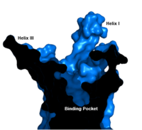
| - | [[Image:Labeled_Binding_Pocket.png|200 px|left|thumb|'''Figure 3. GCGR Binding Pocket.''' A cross-section of the GCGR binding pocket shows its width and depth]]The class B GPCR has the widest and longest <scene name='72/721538/Binding_pocket/1'>binding pocket</scene> of all other classes of GPCRs. The distance between the EC tips of | + | [[Image:Labeled_Binding_Pocket.png|200 px|left|thumb|'''Figure 3. GCGR Binding Pocket.''' A cross-section of the GCGR binding pocket shows its width and depth.]]The class B GPCR has the widest and longest <scene name='72/721538/Binding_pocket/1'>binding pocket</scene> of all other classes of GPCRs. The distance between the EC tips of Helices II and VI and between the tips of Helices III and VII are some of the largest among the GPCRs.<ref name= "Siu 2013"/> As a result, the [http://www.ncbi.nlm.nih.gov/pmc/articles/PMC3820480/bin/nihms495648f2.jpg binding cavity] of GCGR is located deeper inside the receptor, meaning glucagon binds much closer to the cell membrane. |
====Other Unique Structural Features ==== | ====Other Unique Structural Features ==== | ||
| - | An important interface stabilization interaction between Helices I and VII occurs between [https://en.wikipedia.org/wiki/Serine Ser] 152 of Helix I and Ser 390 of Helix VII. Due to their close proximity to one another, they form an important <scene name='72/721537/Ser-ser_hydrogen_bond/3'>hydrogen bond</scene> which stabilizes the structure of GCGR. Mutations to the homologous residues Ser 135 and Ser 392 | + | An important interface stabilization interaction between Helices I and VII occurs between [https://en.wikipedia.org/wiki/Serine Ser] 152 of Helix I and Ser 390 of Helix VII. Due to their close proximity to one another, they form an important <scene name='72/721537/Ser-ser_hydrogen_bond/3'>hydrogen bond</scene> which stabilizes the structure of GCGR. Mutations to the homologous residues Ser 135 and Ser 392 alters receptor signaling in [https://en.wikipedia.org/wiki/Glucagon-like_peptide_1_receptor glucagon-like peptide-1 receptor] (GLP1R). |
===Glucagon Binding=== | ===Glucagon Binding=== | ||
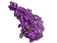
The large, soluble N-terminal extracellular domains (ECD) of GCGR provide initial ligand selectivity with the deep, ligand pocket of the TMD providing secondary recognition.<ref name= "Yang 2015"/> [[Image:ECD_bound_to_glucagon.png|200 px|left|thumb|'''Figure 4. Bound Molecule of Glucagon.''' A molecule of glucagon is shown bound to the GCGR's ECD (shown in magenta)]] The active, or open conformation, is characterized by an intracellular outward movement of <scene name='72/721538/Helix_v_and_vi/1'>helicies V and VI</scene> (breaking hydrogen bonds between <scene name='72/721538/Arg173-ser350_h_bond/1'>Arg173-Ser350</scene> and <scene name='72/721538/Arg173-ser350_h_bond/2'>Glu245-Thr351</scene>)<ref name= "Hollenstein 2014"/> and an extracellular rotation of the ECD until it is almost perpendicular to the membrane surface.<ref name="Lastt"/> While the stalk region of Helix I helps to facilitate the motion of the ECD, intracellular G-protein coupling and extracellular glucagon binding stabilized this active state. In the abscence of glucagon, however, the GCGR adopts a closed conformation in which all three of the extracellular loops of the 7tm (<scene name='72/721538/Ecls/1'>ECL1, ECL2, and ECL3</scene>) can interact with the ECD.<ref name="Lastt"/> In this closed state, the ECD covers the extracellular surface of the 7tm. To transition between states, the ECD rotates and moves down towards the 7tm domain. This transition mechanism is consistent with the "two-domain" binding mechanism of class B GCPRs in which (1) the C-terminus of the ligand first binds to the ECD allowing (2) the N-terminus of the ligand to interact with the 7tm and activate the protein.<ref name= "Hollenstein 2014"/> | The large, soluble N-terminal extracellular domains (ECD) of GCGR provide initial ligand selectivity with the deep, ligand pocket of the TMD providing secondary recognition.<ref name= "Yang 2015"/> [[Image:ECD_bound_to_glucagon.png|200 px|left|thumb|'''Figure 4. Bound Molecule of Glucagon.''' A molecule of glucagon is shown bound to the GCGR's ECD (shown in magenta)]] The active, or open conformation, is characterized by an intracellular outward movement of <scene name='72/721538/Helix_v_and_vi/1'>helicies V and VI</scene> (breaking hydrogen bonds between <scene name='72/721538/Arg173-ser350_h_bond/1'>Arg173-Ser350</scene> and <scene name='72/721538/Arg173-ser350_h_bond/2'>Glu245-Thr351</scene>)<ref name= "Hollenstein 2014"/> and an extracellular rotation of the ECD until it is almost perpendicular to the membrane surface.<ref name="Lastt"/> While the stalk region of Helix I helps to facilitate the motion of the ECD, intracellular G-protein coupling and extracellular glucagon binding stabilized this active state. In the abscence of glucagon, however, the GCGR adopts a closed conformation in which all three of the extracellular loops of the 7tm (<scene name='72/721538/Ecls/1'>ECL1, ECL2, and ECL3</scene>) can interact with the ECD.<ref name="Lastt"/> In this closed state, the ECD covers the extracellular surface of the 7tm. To transition between states, the ECD rotates and moves down towards the 7tm domain. This transition mechanism is consistent with the "two-domain" binding mechanism of class B GCPRs in which (1) the C-terminus of the ligand first binds to the ECD allowing (2) the N-terminus of the ligand to interact with the 7tm and activate the protein.<ref name= "Hollenstein 2014"/> | ||
| - | The [https://en.wikipedia.org/wiki/Residue_(chemistry) residues] in the binding pocket that are in direct contact with the glucagon molecule are [https://en.wikipedia.org/wiki/Chemical_polarity polar] | + | The [https://en.wikipedia.org/wiki/Residue_(chemistry) residues] in the binding pocket that are in direct contact with the glucagon molecule are [https://en.wikipedia.org/wiki/Chemical_polarity polar] or are hydrophobic. The N-terminus of glucagon binds partly with the ECD while the rest of glucagon binds deep into the binding pocket. The [https://en.wikipedia.org/wiki/Amino_acid amino acids] at the N-terminus of the class B 7TM have the ability to form [https://en.wikipedia.org/wiki/Hydrogen_bond hydrogen bonds] and [https://en.wikipedia.org/wiki/Ionic_bonding ionic interactions], which can be seen in the [https://en.wikipedia.org/wiki/Peptide_sequence amino acid sequence] of glucagon (Figure 5). <ref name="Sequence">PMID: 11946536</ref> |
[[Image:Glucagonstructure.png|(|):|360 px|right|thumb|'''Figure 5: Structure of Glucagon:''' The side chains of the residues making up glucagon are depicted. Coloration on the side chains indicate certain [https://en.wikipedia.org/wiki/Atom atoms] that determine the properties the residues hold. The blue indicates a [https://en.wikipedia.org/wiki/Nitrogen nitrogen] atom (hydrophilic properties), the green on the side chains indicates carbon atoms (non-polar hydrophobic properties), and the red coloration indicates an [https://en.wikipedia.org/wiki/Oxygen oxygen] atom (hydrophilic properties). [http://www.rcsb.org/pdb/home/home.do PDB] [http://www.rcsb.org/pdb/explore.do?structureId=1GCN 1GCN] ]] | [[Image:Glucagonstructure.png|(|):|360 px|right|thumb|'''Figure 5: Structure of Glucagon:''' The side chains of the residues making up glucagon are depicted. Coloration on the side chains indicate certain [https://en.wikipedia.org/wiki/Atom atoms] that determine the properties the residues hold. The blue indicates a [https://en.wikipedia.org/wiki/Nitrogen nitrogen] atom (hydrophilic properties), the green on the side chains indicates carbon atoms (non-polar hydrophobic properties), and the red coloration indicates an [https://en.wikipedia.org/wiki/Oxygen oxygen] atom (hydrophilic properties). [http://www.rcsb.org/pdb/home/home.do PDB] [http://www.rcsb.org/pdb/explore.do?structureId=1GCN 1GCN] ]] | ||
| - | + | GCGR regions providing binding affinity for glucagon include the α-helical structure of the <scene name='72/721535/Stalk/1'>stalk</scene>. The α-helical structure of the stalk interacts directly with glucagon, as it extends nearly three helical turns above the membrane. When the alpha helix of the stalk is disrupted, the affinity of glucagon for GCGR decreases with an <scene name='72/721535/Ala135/1'>alanine 135</scene> to proline substitution having significantly lower affinity for glucagon.<ref name= "Siu 2013"/> The high affinity conformation of GCGR is the open conformation, when glucagon can bind (Figure 2). The [https://en.wikipedia.org/wiki/Disulfide disulfide bond] between <scene name='72/721535/Disulfide_bond_notspin_actual/2'>Cys 294 and Cys 224</scene> serves to hold the helices in the proper orientation for binding and stabilizes the open conformation. Additionally, the [https://en.wikipedia.org/wiki/Salt_bridge_%28protein_and_supramolecular%29 salt bridges] between <scene name='72/721535/Salt_bridge_residues/1'>Glu 406, Arg 173, and Arg 346</scene> hold the open conformation together for higher affinity. <ref name="Ligands">PMID: 21542831</ref> | |
| - | <scene name='72/721535/Salt_bridge_residues/1'>Glu 406, Arg 173, and Arg 346</scene> hold the open conformation together for higher affinity. <ref name="Ligands">PMID: 21542831</ref> | + | |
| - | Mutagenesis and photo cross-linking studies determined essential, conserved residues in glucagon and have been <scene name='72/727091/Glucagon_important_residues/2'>labeled and colored</scene> in red.<ref name= "Siu 2013"/> Glucagon residues His 1, Gln 3, Phe 6, and Tyr 10 are critical to successful binding interaction with the GCGR while others are important for structural rigidity. The n-terminus of glucagon ( | + | Mutagenesis and photo cross-linking studies determined essential, conserved residues in glucagon and have been <scene name='72/727091/Glucagon_important_residues/2'>labeled and colored</scene> in red.<ref name= "Siu 2013"/> Glucagon residues His 1, Gln 3, Phe 6, and Tyr 10 are critical to successful binding interaction with the GCGR while others are important for structural rigidity. The n-terminus of glucagon (Figure 5) leads to a protuberance that fits into the deep, interior cavity of the GCGR 7TMD (Figure 3) where four residues reside that play strong roles in ligand binding affinity. There is a <scene name='72/721552/Glucagon_binding_zoomed_in/1'>narrow neck</scene> to the entrance of the cavity, providing a firm anchor during peptide docking (Figure 3). |
==Glucagon Signaling Pathway== | ==Glucagon Signaling Pathway== | ||
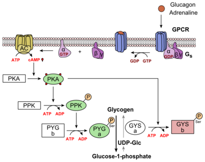
| - | Glucagon binds to the open conformation of GCGR | + | Glucagon binds to the open conformation of GCGR on the [https://en.wikipedia.org/wiki/Cell_membrane plasma membrane]. Glucagon binding to GCGR induces a [https://en.wikipedia.org/wiki/Conformational_change conformational change] in GCGR. This conformation change induces the active state of the protein (Figure 2). The active state of the protein exchanges a [https://en.wikipedia.org/wiki/Guanosine_diphosphate guanosine diphosphate (GDP]) for [https://en.wikipedia.org/wiki/Guanosine_triphosphate guanosine triphosphate (GTP)] that is bound to the [https://en.wikipedia.org/wiki/G_alpha_subunit alpha subunit]. With the GTP in place, the activated alpha subunit dissociates from the [https://en.wikipedia.org/wiki/Heterotrimeric_G_protein heterotrimeric G protein's]beta and gamma subunits. Following dissociation, the alpha subunit can activate [https://en.wikipedia.org/wiki/Adenylyl_cyclase adenylate cyclase]. Activated adenylate cyclase, catalyzes the conversion of [https://en.wikipedia.org/wiki/Adenosine_triphosphate adenosine triphosphate (ATP)] into [https://en.wikipedia.org/wiki/Cyclic_adenosine_monophosphate cyclic adenosine monophosphate (cAMP)]. cAMP then serves as a secondary messenger to activate, through allosteric binding, [https://en.wikipedia.org/wiki/Protein_kinase_A cAMP dependent protein kinase A (PKA)]. PKA activates via [https://en.wikipedia.org/wiki/Phosphorylation phosphorylation] the [https://en.wikipedia.org/wiki/Phosphorylase_kinase phosphorylase b kinase]. The phosphorylase b kinase phosphorylates [https://en.wikipedia.org/wiki/Glycogen_phosphorylase glycogen phosphorylase b] to convert to the active form, phosphorylase a. Phosphorylase a finally catalyzes the release of [https://en.wikipedia.org/wiki/Glucose_1-phosphate glucose-1-phosphate] into the bloodstream from glycogen [https://en.wikipedia.org/wiki/Polymer polymers] (Figure 6). |
[[Image:Glucagon_Pathway.png|(|):|400 px|center|thumb|'''Figure 6: [https://en.wikipedia.org/wiki/Glucagon Metabolic Regulation of Glycogen by Glucagon.]'''Depicted is the visualization of the glucagon signaling pathway through the GCGR. The location of the GCGR, the release of the alpha subunit from the beta and gamma subunits, and the enzyme cascade to result in the releasing of glucose are depicted. Abbreviations for the enzymes in the cascade include- PPK: phosphorylase kinase; PYG b: glycogen phosphorylase b; PYG a: glycogen phosphorylase a.]] | [[Image:Glucagon_Pathway.png|(|):|400 px|center|thumb|'''Figure 6: [https://en.wikipedia.org/wiki/Glucagon Metabolic Regulation of Glycogen by Glucagon.]'''Depicted is the visualization of the glucagon signaling pathway through the GCGR. The location of the GCGR, the release of the alpha subunit from the beta and gamma subunits, and the enzyme cascade to result in the releasing of glucose are depicted. Abbreviations for the enzymes in the cascade include- PPK: phosphorylase kinase; PYG b: glycogen phosphorylase b; PYG a: glycogen phosphorylase a.]] | ||
==Clinical relevance == | ==Clinical relevance == | ||
| - | Because GCGR can interact with multiple types of G protein subfamilies, discovering small molecule inhibitors could lead to a wide range of focused therapies.<ref name= "Weston 2015"/> Blocking conformations that favor interaction with specific G proteins could allow the knockdown of targeted signal pathways. For example, GCGR | + | Because GCGR can interact with multiple types of G protein subfamilies, discovering small molecule inhibitors could lead to a wide range of focused therapies.<ref name= "Weston 2015"/> Blocking conformations that favor interaction with specific G proteins could allow the knockdown of targeted signal pathways. For example, GCGR interacts with inhibitory Gαi proteins that antagonize cAMP production.<ref name= "Weston 2015"/> Finding an agonist for this pathway could be used to treat diabetes mellitus. Current attempts to target the GCGR have however been relatively unsuccessful. Small molecule modulators have been reported with enhanced pharmaceutical regulation, but the progress has been modest.<ref name= "Kazda 2015">DOI: 10.1021/jm058026u</ref> Encouraging results have recently come from Eli Lilly and Company who have been testing a small molecule antagonist of the GCGR (LY2409021) in phase two trials, providing hope for more specific control of diabetes mellitus.<ref name= "Kazda 2015">DOI: 10.2337/dc15-1643</ref> In addition to diabetes mellitus, future development of signal bias modulators promise to provide focused therapies for obesity, heart disease, hypertension, and cancer. |
| - | + | ||
| - | + | ||
==See Also== | ==See Also== | ||
| Line 59: | Line 56: | ||
[https://en.wikipedia.org/wiki/G_protein%E2%80%93coupled_receptor G protein-coupled receptors] | [https://en.wikipedia.org/wiki/G_protein%E2%80%93coupled_receptor G protein-coupled receptors] | ||
| - | == | + | ==Proteopedia Resources== |
| - | + | [http://proteopedia.org/wiki/index.php/Glucagon Glucagon] | |
| + | |||
| + | [http://proteopedia.org/wiki/index.php/Category:Glucagon_receptor Category:Glucagon Receptor] | ||
| + | |||
| + | [http://proteopedia.org/wiki/index.php/User:R._Jeremy_Johnson/CH462:Biochemistry_II_Butler_University Butler University Proteopedia Pages] | ||
</StructureSection> | </StructureSection> | ||
| + | |||
| + | ==Student Contributors== | ||
| + | Steven Bennett | ||
| + | |||
| + | Sydney Caskey | ||
| + | |||
| + | Alexis Coulis | ||
| + | |||
| + | Olivia Murfield | ||
| + | |||
| + | Allie Paton | ||
| + | |||
| + | Dean Williams | ||
Revision as of 14:49, 20 June 2016
Glucagon G protein-coupled receptor
Student Contributors
Steven Bennett
Sydney Caskey
Alexis Coulis
Olivia Murfield
Allie Paton
Dean Williams


