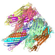
Pore forming toxin, α-hemolysin
From Proteopedia
m (edits text to add cylinder representing membrane location) |
m (delete the test scene call) |
||
| Line 35: | Line 35: | ||
<!-- Note that <scene name='7ahl/7ahlopm/2a'> used to be the best OPM scene but finally seems Jmol straightened out and I could remake scene here by copying state script from that scene in page since couldn't get in scene authoring tools. Remade better. See <scene name='40/404876/Opm_withcyl/2' --> | <!-- Note that <scene name='7ahl/7ahlopm/2a'> used to be the best OPM scene but finally seems Jmol straightened out and I could remake scene here by copying state script from that scene in page since couldn't get in scene authoring tools. Remade better. See <scene name='40/404876/Opm_withcyl/2' --> | ||
To help give a better idea of how α-hemolysin interacts with the membrane lipid bilayer, <scene name='40/404876/Opm_withcyl/3'>a slab representative of the hydrophobic core of the lipid bilayer</scene> as calculated by the [http://opm.phar.umich.edu/protein.php?pdbid=7ahl Orientations of Proteins in Membranes database](University of Michigan, USA) is shown as a cylinder with the red patch ndicating the boundary of the hydrophobic core closet to the outside of the cell and the blue patch indicating the boundary closest to the inside of the cell. | To help give a better idea of how α-hemolysin interacts with the membrane lipid bilayer, <scene name='40/404876/Opm_withcyl/3'>a slab representative of the hydrophobic core of the lipid bilayer</scene> as calculated by the [http://opm.phar.umich.edu/protein.php?pdbid=7ahl Orientations of Proteins in Membranes database](University of Michigan, USA) is shown as a cylinder with the red patch ndicating the boundary of the hydrophobic core closet to the outside of the cell and the blue patch indicating the boundary closest to the inside of the cell. | ||
| - | <scene name='40/404876/Opm_withcyl/3'>test cylinder</scene> | ||
| | ||
Revision as of 00:48, 10 July 2016
α-Hemolysin from Staphlococcus aureus is a pore-forming toxin made of seven repeats of an identical monomer arranged in a ring. The structural basis of the toxic activity was readily revealed when the structure of the ring was solved.
Contents |
ALPHA-HEMOLYSIN FROM STAPHYLOCOCCUS AUREUS
The structure of the Staphylococcus aureus alpha-hemolysin pore has been determined to 1.9 A resolution. Contained within the mushroom-shaped homo-oligomeric heptamer is a solvent-filled channel, 100 A in length, that runs along the sevenfold axis and ranges from 14 A to 46 A in diameter. The lytic, transmembrane domain comprises the lower half of a 14-strand antiparallel beta barrel, to which each protomer contributes two beta strands, each 65 A long. The interior of the beta barrel is primarily hydrophilic, and the exterior has a hydrophobic belt 28 A wide. The structure proves the heptameric subunit stoichiometry of the alpha-hemolysin oligomer, shows that a glycine-rich and solvent-exposed region of a water-soluble protein can self-assemble to form a transmembrane pore of defined structure, and provides insight into the principles of membrane interaction and transport activity of beta barrel pore-forming toxins.
Structure of staphylococcal alpha-hemolysin, a heptameric transmembrane pore., Song L, Hobaugh MR, Shustak C, Cheley S, Bayley H, Gouaux JE, Science. 1996 Dec 13;274(5294):1859-66. PMID:8943190
From MEDLINE®/PubMed®, a database of the U.S. National Library of Medicine.
Background
Staphylococcus aureus is often pathogenic to humans and relies on several virulence factors in the course of the infections. α-hemolysin is one such virulencce factor that forms pores in target cells[1], similar to other hemolsyins.
About this Structure
| |||||||||||||
|
| |||||||||||||
Use of α-hemolysin in DNA sequencing technology
In 1996, researchers from Harvard University and the University of California at Santa Cruz, including Daniel Branton, David Deamer and John Kasianowicz showed that the pore structure formed by α-hemolysin could be used to pass a single strand of DNA through a nanopore [2]. This was later linked to an exonuclease and a detection mechanism for residency of individual nucleotides in the base of pore to ultimately form the basis of sequencing technology finally offered by Oxford Nanopore, although the specifics of the current generation of the technology may be different [3][4].
PDB entry
7ahl is a 7 chain structure of sequences from Staphylococcus aureus. Full crystallographic information is available from OCA.
Reference for the structure
- Song L, Hobaugh MR, Shustak C, Cheley S, Bayley H, Gouaux JE. Structure of staphylococcal alpha-hemolysin, a heptameric transmembrane pore. Science. 1996 Dec 13;274(5294):1859-66. PMID:8943190
Notes and Literature References
- ↑ Menestrina G, Dalla Serra M, Comai M, Coraiola M, Viero G, Werner S, Colin DA, Monteil H, Prevost G. Ion channels and bacterial infection: the case of beta-barrel pore-forming protein toxins of Staphylococcus aureus. FEBS Lett. 2003 Sep 18;552(1):54-60. PMID:12972152
- ↑ Kasianowicz JJ, Brandin E, Branton D, Deamer DW. Characterization of individual polynucleotide molecules using a membrane channel. Proc Natl Acad Sci U S A. 1996 Nov 26;93(24):13770-3. PMID:8943010
- ↑ Nanopore sequencing: Towards the 15-minute genome. The Economist. Technology Quarterly: Q1 2011. Mar 10th 2011. http://www.economist.com/node/18304268
- ↑ Pennisi E. Genome sequencing. Search for pore-fection. Science. 2012 May 4;336(6081):534-7. PMID:22556226 doi:10.1126/science.336.6081.534
3D structures of hemolysin
see, Hemolysin
See Also
- Hemolysin
- 1lkf,2lkf & 3lkf, Leukocidin F (Hlg B) from Staphylococcus aureus
- 1pvl, the F component of Panton-Valentine leucocidin from Staphylococcus aureus
- 3fy3
- Membrane proteins
- Membrane Channels & Pumps
Additional Literature and Resources
- For additional information, see: Toxins
- David Briggs's Blog on α-hemolsyin, which served as a template for the original adaptation of 7ahl to this topic page.
- Pore-forming toxin at Wikipedia
- Page on Bacterial Toxin α-Hemolysin by Aleksei Aksimentiev and Klaus Schulten
Page started with original page seeded by OCA on Mon Feb 16 13:00:23 2009 for 7ahl

