1lpa
From Proteopedia
| Line 4: | Line 4: | ||
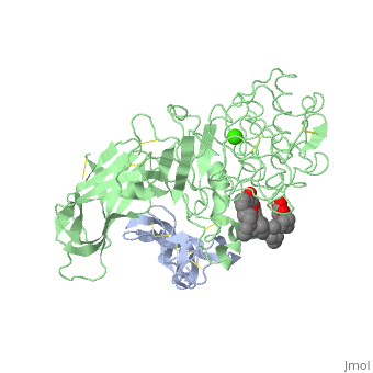
|PDB= 1lpa |SIZE=350|CAPTION= <scene name='initialview01'>1lpa</scene>, resolution 3.04Å | |PDB= 1lpa |SIZE=350|CAPTION= <scene name='initialview01'>1lpa</scene>, resolution 3.04Å | ||
|SITE= | |SITE= | ||
| - | |LIGAND= <scene name='pdbligand=BNG:B-NONYLGLUCOSIDE'>BNG</scene>, <scene name='pdbligand=CA:CALCIUM+ION'>CA</scene> | + | |LIGAND= <scene name='pdbligand=BNG:B-NONYLGLUCOSIDE'>BNG</scene>, <scene name='pdbligand=CA:CALCIUM+ION'>CA</scene>, <scene name='pdbligand=PLC:DIUNDECYL+PHOSPHATIDYL+CHOLINE'>PLC</scene> |
| - | |ACTIVITY= [http://en.wikipedia.org/wiki/Triacylglycerol_lipase Triacylglycerol lipase], with EC number [http://www.brenda-enzymes.info/php/result_flat.php4?ecno=3.1.1.3 3.1.1.3] | + | |ACTIVITY= <span class='plainlinks'>[http://en.wikipedia.org/wiki/Triacylglycerol_lipase Triacylglycerol lipase], with EC number [http://www.brenda-enzymes.info/php/result_flat.php4?ecno=3.1.1.3 3.1.1.3] </span> |
|GENE= | |GENE= | ||
| + | |DOMAIN= | ||
| + | |RELATEDENTRY= | ||
| + | |RESOURCES=<span class='plainlinks'>[http://oca.weizmann.ac.il/oca-docs/fgij/fg.htm?mol=1lpa FirstGlance], [http://oca.weizmann.ac.il/oca-bin/ocaids?id=1lpa OCA], [http://www.ebi.ac.uk/pdbsum/1lpa PDBsum], [http://www.rcsb.org/pdb/explore.do?structureId=1lpa RCSB]</span> | ||
}} | }} | ||
| Line 14: | Line 17: | ||
==Overview== | ==Overview== | ||
The three-dimensional structure of the lipase-procolipase complex, co-crystallized with mixed micelles of phosphatidylcholine and bile salt, has been determined at 3 A resolution by X-ray crystallography. The lid, a surface helix covering the catalytic triad of lipase, adopts a totally different conformation which allows phospholipid to bind to the enzyme's active site. The open lid is an essential component of the active site and interacts with procolipase. Together they form the lipid-water interface binding site. This reorganization of the lid structure provokes a second drastic conformational change in an active site loop, which in its turn creates the oxyanion hole (induced fit). | The three-dimensional structure of the lipase-procolipase complex, co-crystallized with mixed micelles of phosphatidylcholine and bile salt, has been determined at 3 A resolution by X-ray crystallography. The lid, a surface helix covering the catalytic triad of lipase, adopts a totally different conformation which allows phospholipid to bind to the enzyme's active site. The open lid is an essential component of the active site and interacts with procolipase. Together they form the lipid-water interface binding site. This reorganization of the lid structure provokes a second drastic conformational change in an active site loop, which in its turn creates the oxyanion hole (induced fit). | ||
| - | |||
| - | ==Disease== | ||
| - | Known disease associated with this structure: Pancreatic lipase deficiency OMIM:[[http://www.ncbi.nlm.nih.gov/entrez/dispomim.cgi?id=246600 246600]] | ||
==About this Structure== | ==About this Structure== | ||
| Line 30: | Line 30: | ||
[[Category: Egloff, M P.]] | [[Category: Egloff, M P.]] | ||
[[Category: Tilbeurgh, H Van.]] | [[Category: Tilbeurgh, H Van.]] | ||
| - | [[Category: BNG]] | ||
| - | [[Category: CA]] | ||
| - | [[Category: PLC]] | ||
[[Category: hydrolase(carboxylic esterase)]] | [[Category: hydrolase(carboxylic esterase)]] | ||
| - | ''Page seeded by [http://oca.weizmann.ac.il/oca OCA ] on | + | ''Page seeded by [http://oca.weizmann.ac.il/oca OCA ] on Sun Mar 30 22:05:37 2008'' |
Revision as of 19:05, 30 March 2008
| |||||||
| , resolution 3.04Å | |||||||
|---|---|---|---|---|---|---|---|
| Ligands: | , , | ||||||
| Activity: | Triacylglycerol lipase, with EC number 3.1.1.3 | ||||||
| Resources: | FirstGlance, OCA, PDBsum, RCSB | ||||||
| Coordinates: | save as pdb, mmCIF, xml | ||||||
INTERFACIAL ACTIVATION OF THE LIPASE-PROCOLIPASE COMPLEX BY MIXED MICELLES REVEALED BY X-RAY CRYSTALLOGRAPHY
Overview
The three-dimensional structure of the lipase-procolipase complex, co-crystallized with mixed micelles of phosphatidylcholine and bile salt, has been determined at 3 A resolution by X-ray crystallography. The lid, a surface helix covering the catalytic triad of lipase, adopts a totally different conformation which allows phospholipid to bind to the enzyme's active site. The open lid is an essential component of the active site and interacts with procolipase. Together they form the lipid-water interface binding site. This reorganization of the lid structure provokes a second drastic conformational change in an active site loop, which in its turn creates the oxyanion hole (induced fit).
About this Structure
1LPA is a Protein complex structure of sequences from Homo sapiens and Sus scrofa. Full crystallographic information is available from OCA.
Reference
Interfacial activation of the lipase-procolipase complex by mixed micelles revealed by X-ray crystallography., van Tilbeurgh H, Egloff MP, Martinez C, Rugani N, Verger R, Cambillau C, Nature. 1993 Apr 29;362(6423):814-20. PMID:8479519
Page seeded by OCA on Sun Mar 30 22:05:37 2008
