This old version of Proteopedia is provided for student assignments while the new version is undergoing repairs. Content and edits done in this old version of Proteopedia after March 1, 2026 will eventually be lost when it is retired in about June of 2026.
Apply for new accounts at the new Proteopedia. Your logins will work in both the old and new versions.
Plasminogen
From Proteopedia
(Difference between revisions)
| Line 10: | Line 10: | ||
== Structural highlights == | == Structural highlights == | ||
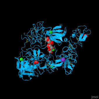
<scene name='46/465445/Cv/2'>PLN contains 7 domains</scene> which are: N-terminal, C-terminal serine protease catalytic domain and 5 kringle domains of ca. 80 residues. The <scene name='46/465445/Cv/3'>kringle domain folds into a large loop containing 3 disulfide bonds</scene>, ''e.g.'' <scene name='46/465445/Cv/4'>Kringle 4</scene>. The kringle domain is important in protein-protein interaction with blood coagulation factors.<ref>PMID:22832192</ref>. | <scene name='46/465445/Cv/2'>PLN contains 7 domains</scene> which are: N-terminal, C-terminal serine protease catalytic domain and 5 kringle domains of ca. 80 residues. The <scene name='46/465445/Cv/3'>kringle domain folds into a large loop containing 3 disulfide bonds</scene>, ''e.g.'' <scene name='46/465445/Cv/4'>Kringle 4</scene>. The kringle domain is important in protein-protein interaction with blood coagulation factors.<ref>PMID:22832192</ref>. | ||
| + | *<scene name='46/465445/Cv/6'>K ion coordination site</scene>. Water molecules shown as red spheres. | ||
| + | *<scene name='46/465445/Cv/7'>Cl ion coordination site I</scene>. | ||
| + | *<scene name='46/465445/Cv/8'>Cl ion coordination site II</scene>. | ||
| + | *<scene name='46/465445/Cv/9'>Cl ion coordination site III</scene>. | ||
</StructureSection> | </StructureSection> | ||
== 3D Structures of plasminogen == | == 3D Structures of plasminogen == | ||
Revision as of 09:29, 15 August 2016
| |||||||||||
3D Structures of plasminogen
Updated on 15-August-2016
References
- ↑ Goldenberg DT, Giblin FJ, Cheng M, Chintala SK, Trese MT, Drenser KA, Ruby AJ. Posterior vitreous detachment with microplasmin alters the retinal penetration of intravitreal bevacizumab (Avastin) in rabbit eyes. Retina. 2011 Feb;31(2):393-400. doi: 10.1097/IAE.0b013e3181e586b2. PMID:21099453 doi:http://dx.doi.org/10.1097/IAE.0b013e3181e586b2
- ↑ Mehta R, Shapiro AD. Plasminogen deficiency. Haemophilia. 2008 Nov;14(6):1261-8. doi: 10.1111/j.1365-2516.2008.01825.x. PMID:19141167 doi:http://dx.doi.org/10.1111/j.1365-2516.2008.01825.x
- ↑ Law RH, Caradoc-Davies T, Cowieson N, Horvath AJ, Quek AJ, Encarnacao JA, Steer D, Cowan A, Zhang Q, Lu BG, Pike RN, Smith AI, Coughlin PB, Whisstock JC. The X-ray crystal structure of full-length human plasminogen. Cell Rep. 2012 Mar 29;1(3):185-90. doi: 10.1016/j.celrep.2012.02.012. Epub 2012, Mar 8. PMID:22832192 doi:10.1016/j.celrep.2012.02.012
