This old version of Proteopedia is provided for student assignments while the new version is undergoing repairs. Content and edits done in this old version of Proteopedia after March 1, 2026 will eventually be lost when it is retired in about June of 2026.
Apply for new accounts at the new Proteopedia. Your logins will work in both the old and new versions.
Phosphomannomutase
From Proteopedia
(Difference between revisions)
| Line 7: | Line 7: | ||
== Structural highlights == | == Structural highlights == | ||
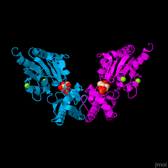
| - | The PMM structure contains a <scene name='48/481638/Cv/ | + | The PMM structure contains a <scene name='48/481638/Cv/6'>core domain and a cap domain</scene>. It is known that in order for the substrate to bind, the cap must dissociate from the core domain and then re-associate to seal the active site from solvent molecules. The <scene name='48/481638/Cv/4'>active site of PMM is located at the interface of the core and cap domains of the molecule</scene><ref>PMID:16540464</ref>. Water molecules shown as red spheres. |
</StructureSection> | </StructureSection> | ||
Revision as of 09:40, 4 September 2016
| |||||||||||
3D Structures of phosphomannomutase
Updated on 04-September-2016
References
- ↑ Matthijs G, Schollen E, Pardon E, Veiga-Da-Cunha M, Jaeken J, Cassiman JJ, Van Schaftingen E. Mutations in PMM2, a phosphomannomutase gene on chromosome 16p13, in carbohydrate-deficient glycoprotein type I syndrome (Jaeken syndrome). Nat Genet. 1997 May;16(1):88-92. PMID:9140401 doi:10.1038/ng0597-88
- ↑ Silvaggi NR, Zhang C, Lu Z, Dai J, Dunaway-Mariano D, Allen KN. The X-ray crystal structures of human alpha-phosphomannomutase 1 reveal the structural basis of congenital disorder of glycosylation type 1a. J Biol Chem. 2006 May 26;281(21):14918-26. Epub 2006 Mar 15. PMID:16540464 doi:http://dx.doi.org/10.1074/jbc.M601505200
