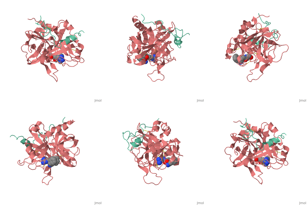
1ppb
From Proteopedia
| Line 4: | Line 4: | ||
|PDB= 1ppb |SIZE=350|CAPTION= <scene name='initialview01'>1ppb</scene>, resolution 1.92Å | |PDB= 1ppb |SIZE=350|CAPTION= <scene name='initialview01'>1ppb</scene>, resolution 1.92Å | ||
|SITE= | |SITE= | ||
| - | |LIGAND= <scene name='pdbligand=CH2:METHYLENE GROUP'>CH2</scene> | + | |LIGAND= <scene name='pdbligand=CH2:METHYLENE+GROUP'>CH2</scene> |
| - | |ACTIVITY= [http://en.wikipedia.org/wiki/Thrombin Thrombin], with EC number [http://www.brenda-enzymes.info/php/result_flat.php4?ecno=3.4.21.5 3.4.21.5] | + | |ACTIVITY= <span class='plainlinks'>[http://en.wikipedia.org/wiki/Thrombin Thrombin], with EC number [http://www.brenda-enzymes.info/php/result_flat.php4?ecno=3.4.21.5 3.4.21.5] </span> |
|GENE= | |GENE= | ||
| + | |DOMAIN= | ||
| + | |RELATEDENTRY= | ||
| + | |RESOURCES=<span class='plainlinks'>[http://oca.weizmann.ac.il/oca-docs/fgij/fg.htm?mol=1ppb FirstGlance], [http://oca.weizmann.ac.il/oca-bin/ocaids?id=1ppb OCA], [http://www.ebi.ac.uk/pdbsum/1ppb PDBsum], [http://www.rcsb.org/pdb/explore.do?structureId=1ppb RCSB]</span> | ||
}} | }} | ||
| Line 14: | Line 17: | ||
==Overview== | ==Overview== | ||
A stoichiometric complex formed between human alpha-thrombin and D-Phe-Pro-Arg chloromethylketone was crystallized in an orthorhombic crystal form. Orientation and position of a starting model derived from homologous modelling were determined by Patterson search methods. The thrombin model was completed in a cyclic modelling-crystallographic refinement procedure to a final R-value of 0.171 for X-ray data to 1.92 A. The structure is in full agreement with published cDNA sequence data. The A-chain, ordered only in its central part, is positioned along the molecular surface opposite to the active site. The B-chain exhibits the characteristic polypeptide fold of trypsin-like proteinases. Several extended insertions form, however, large protuberances; most important for interaction with macromolecular substrates is the characteristic thrombin loop around Tyr60A-Pro60B-Pro60C-Trp60D (chymotrypsinogen numbering) and the enlarged loop around the unique Trp148. The former considerably restricts the active site cleft and seems likely to be responsible for poor binding of most natural proteinase inhibitors to thrombin. The exceptional specificity of D-Phe-Pro-Arg chloromethylketone can be explained by a hydrophobic cage formed by Ile174, Trp215, Leu99, His57, Tyr60A and Trp60D. The narrow active site cleft, with a more polar base and hydrophobic rims, extends towards the arginine-rich surface of loop Lys70-Glu80 that probably represents part of the anionic binding region for hirudin and fibrinogen. | A stoichiometric complex formed between human alpha-thrombin and D-Phe-Pro-Arg chloromethylketone was crystallized in an orthorhombic crystal form. Orientation and position of a starting model derived from homologous modelling were determined by Patterson search methods. The thrombin model was completed in a cyclic modelling-crystallographic refinement procedure to a final R-value of 0.171 for X-ray data to 1.92 A. The structure is in full agreement with published cDNA sequence data. The A-chain, ordered only in its central part, is positioned along the molecular surface opposite to the active site. The B-chain exhibits the characteristic polypeptide fold of trypsin-like proteinases. Several extended insertions form, however, large protuberances; most important for interaction with macromolecular substrates is the characteristic thrombin loop around Tyr60A-Pro60B-Pro60C-Trp60D (chymotrypsinogen numbering) and the enlarged loop around the unique Trp148. The former considerably restricts the active site cleft and seems likely to be responsible for poor binding of most natural proteinase inhibitors to thrombin. The exceptional specificity of D-Phe-Pro-Arg chloromethylketone can be explained by a hydrophobic cage formed by Ile174, Trp215, Leu99, His57, Tyr60A and Trp60D. The narrow active site cleft, with a more polar base and hydrophobic rims, extends towards the arginine-rich surface of loop Lys70-Glu80 that probably represents part of the anionic binding region for hirudin and fibrinogen. | ||
| - | |||
| - | ==Disease== | ||
| - | Known diseases associated with this structure: Dysprothrombinemia OMIM:[[http://www.ncbi.nlm.nih.gov/entrez/dispomim.cgi?id=176930 176930]], Hyperprothrombinemia OMIM:[[http://www.ncbi.nlm.nih.gov/entrez/dispomim.cgi?id=176930 176930]], Hypoprothrombinemia OMIM:[[http://www.ncbi.nlm.nih.gov/entrez/dispomim.cgi?id=176930 176930]] | ||
==About this Structure== | ==About this Structure== | ||
| Line 27: | Line 27: | ||
[[Category: Thrombin]] | [[Category: Thrombin]] | ||
[[Category: Bode, W.]] | [[Category: Bode, W.]] | ||
| - | [[Category: CH2]] | ||
[[Category: hydrolase(serine proteinase)]] | [[Category: hydrolase(serine proteinase)]] | ||
| - | ''Page seeded by [http://oca.weizmann.ac.il/oca OCA ] on | + | ''Page seeded by [http://oca.weizmann.ac.il/oca OCA ] on Sun Mar 30 23:02:41 2008'' |
Revision as of 20:02, 30 March 2008
| |||||||
| , resolution 1.92Å | |||||||
|---|---|---|---|---|---|---|---|
| Ligands: | |||||||
| Activity: | Thrombin, with EC number 3.4.21.5 | ||||||
| Resources: | FirstGlance, OCA, PDBsum, RCSB | ||||||
| Coordinates: | save as pdb, mmCIF, xml | ||||||
THE REFINED 1.9 ANGSTROMS CRYSTAL STRUCTURE OF HUMAN ALPHA-THROMBIN: INTERACTION WITH D-PHE-PRO-ARG CHLOROMETHYLKETONE AND SIGNIFICANCE OF THE TYR-PRO-PRO-TRP INSERTION SEGMENT
Overview
A stoichiometric complex formed between human alpha-thrombin and D-Phe-Pro-Arg chloromethylketone was crystallized in an orthorhombic crystal form. Orientation and position of a starting model derived from homologous modelling were determined by Patterson search methods. The thrombin model was completed in a cyclic modelling-crystallographic refinement procedure to a final R-value of 0.171 for X-ray data to 1.92 A. The structure is in full agreement with published cDNA sequence data. The A-chain, ordered only in its central part, is positioned along the molecular surface opposite to the active site. The B-chain exhibits the characteristic polypeptide fold of trypsin-like proteinases. Several extended insertions form, however, large protuberances; most important for interaction with macromolecular substrates is the characteristic thrombin loop around Tyr60A-Pro60B-Pro60C-Trp60D (chymotrypsinogen numbering) and the enlarged loop around the unique Trp148. The former considerably restricts the active site cleft and seems likely to be responsible for poor binding of most natural proteinase inhibitors to thrombin. The exceptional specificity of D-Phe-Pro-Arg chloromethylketone can be explained by a hydrophobic cage formed by Ile174, Trp215, Leu99, His57, Tyr60A and Trp60D. The narrow active site cleft, with a more polar base and hydrophobic rims, extends towards the arginine-rich surface of loop Lys70-Glu80 that probably represents part of the anionic binding region for hirudin and fibrinogen.
About this Structure
1PPB is a Protein complex structure of sequences from Homo sapiens. The following page contains interesting information on the relation of 1PPB with [Thrombin]. Full crystallographic information is available from OCA.
Reference
The refined 1.9 A crystal structure of human alpha-thrombin: interaction with D-Phe-Pro-Arg chloromethylketone and significance of the Tyr-Pro-Pro-Trp insertion segment., Bode W, Mayr I, Baumann U, Huber R, Stone SR, Hofsteenge J, EMBO J. 1989 Nov;8(11):3467-75. PMID:2583108
Page seeded by OCA on Sun Mar 30 23:02:41 2008

