We apologize for Proteopedia being slow to respond. For the past two years, a new implementation of Proteopedia has been being built. Soon, it will replace this 18-year old system. All existing content will be moved to the new system at a date that will be announced here.
Urease accessory protein
From Proteopedia
(Difference between revisions)
| Line 6: | Line 6: | ||
== Structural highlights == | == Structural highlights == | ||
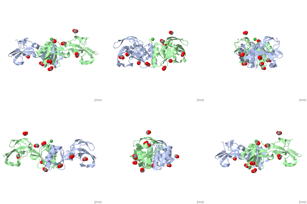
| + | The Ni+2 ion coordinates six ligands which include 3 His residues belonging to the two monomers, Glu from a symmetry-related monomer, water molecule and an unidentified ligand which may be a His from an unobserved part of the chain<ref>PMID:22010876</ref> | ||
</StructureSection> | </StructureSection> | ||
Revision as of 08:13, 8 December 2016
| |||||||||||
3D structures of urease accessory protein
Updated on 08-December-2016
References
- ↑ Witte CP, Isidore E, Tiller SA, Davies HV, Taylor MA. Functional characterisation of urease accessory protein G (ureG) from potato. Plant Mol Biol. 2001 Jan;45(2):169-79. PMID:11289508
- ↑ Banaszak K, Martin-Diaconescu V, Bellucci M, Zambelli B, Rypniewski W, Maroney MJ, Ciurli S. Crystallographic and X-ray absorption spectroscopic characterization of Helicobacter pylori UreE bound to Ni2+ and Zn2+ reveal a role for the disordered C-terminal arm in metal trafficking. Biochem J. 2011 Oct 20. PMID:22010876 doi:10.1042/BJ20111659

