We apologize for Proteopedia being slow to respond. For the past two years, a new implementation of Proteopedia has been being built. Soon, it will replace this 18-year old system. All existing content will be moved to the new system at a date that will be announced here.
P53
From Proteopedia
(Difference between revisions)
| Line 35: | Line 35: | ||
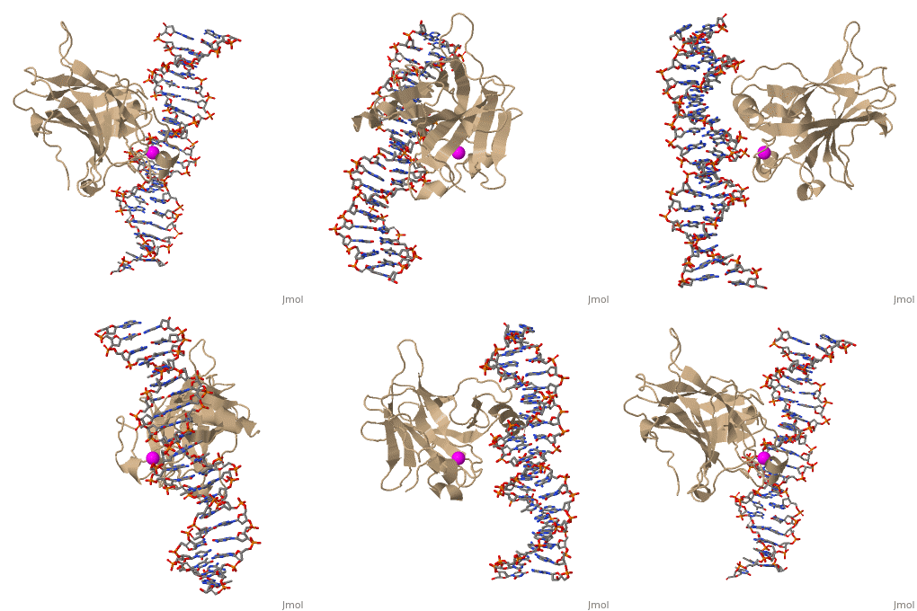
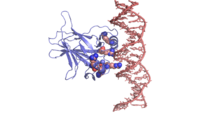
In most cases of human cancer, p53 mutations have been observed. Most of the p53 mutations that may result in cancer are found in and around the DNA-binding surface of the protein. The most common mutation changes can be seen in a close up view of the <scene name='26/26327/B_chain_and_dna/5'>DNA binding domain with DNA </scene> color coded N to C (in rainbow colors), with the amino <scene name='26/26327/B_chain_and_dna/4'>R248</scene> (as space filling spheres) interacting with DNA. When mutated to another amino acid, this interaction is lost. Other residues associated with cancer-causing mutations are arginine <scene name='26/26327/B_chain_and_dna/7'>175, 249, 273, 282 and glycine 245</scene>. Key residues associated with mutations are represented by magenta spheres. | In most cases of human cancer, p53 mutations have been observed. Most of the p53 mutations that may result in cancer are found in and around the DNA-binding surface of the protein. The most common mutation changes can be seen in a close up view of the <scene name='26/26327/B_chain_and_dna/5'>DNA binding domain with DNA </scene> color coded N to C (in rainbow colors), with the amino <scene name='26/26327/B_chain_and_dna/4'>R248</scene> (as space filling spheres) interacting with DNA. When mutated to another amino acid, this interaction is lost. Other residues associated with cancer-causing mutations are arginine <scene name='26/26327/B_chain_and_dna/7'>175, 249, 273, 282 and glycine 245</scene>. Key residues associated with mutations are represented by magenta spheres. | ||
| - | ---- | ||
| - | [[Image:P53_surface_charge.png | left | 400 px | thumb]] | ||
| - | {{Clear}} | ||
=='''Surface charge of the DNA binding domain'''== | =='''Surface charge of the DNA binding domain'''== | ||
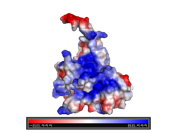
| - | The figure at the left shows the surface charge of the p53 DNA-binding domain. It is rich in arginine amino acids that interact with DNA, and this causes its surface to be positively charged. This domain recognizes specific regulatory sites on the DNA. The flexible structure of p53 allows it to bind to many different variants of binding sites, allowing it to regulate transcription at many places in the genome. | + | [[Image:P53_surface_charge.png | left | 250 px | thumb]]The figure at the left shows the surface charge of the p53 DNA-binding domain )Charged: red - negative; blue - positive). It is rich in arginine amino acids that interact with DNA, and this causes its surface to be positively charged. This domain recognizes specific regulatory sites on the DNA. The flexible structure of p53 allows it to bind to many different variants of binding sites, allowing it to regulate transcription at many places in the genome. |
Revision as of 11:17, 14 January 2017
| |||||||||||
3D structures of p53 (Updated on 14-January-2017)
Additional Resources
References
- ↑ Oren M. Decision making by p53: life, death and cancer. Cell Death Differ. 2003 Apr;10(4):431-42. PMID:12719720 doi:http://dx.doi.org/10.1038/sj.cdd.4401183
- ↑ Oren M, Rotter V. Mutant p53 gain-of-function in cancer. Cold Spring Harb Perspect Biol. 2010 Feb;2(2):a001107. doi:, 10.1101/cshperspect.a001107. PMID:20182618 doi:http://dx.doi.org/10.1101/cshperspect.a001107
Proteopedia Page Contributors and Editors (what is this?)
Joel L. Sussman, Michal Harel, Eran Hodis, Mary Ball, Alexander Berchansky, David Canner


