User:Camille Zumstein/Sandbox
From Proteopedia
(Difference between revisions)
| Line 14: | Line 14: | ||
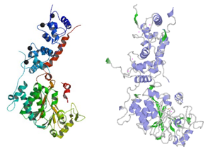
<scene name='75/750223/Jw_caln_sec_strcutur/1'>Calcineurin</scene> is a heterodimeric Protein that consits of two subunits. They are called catalytic and regulatory subunit. Four molecules of Calcineurin form an asymmetric complex which leads to a total molecular weight of 370 kDa. | <scene name='75/750223/Jw_caln_sec_strcutur/1'>Calcineurin</scene> is a heterodimeric Protein that consits of two subunits. They are called catalytic and regulatory subunit. Four molecules of Calcineurin form an asymmetric complex which leads to a total molecular weight of 370 kDa. | ||
| - | The structure presented in this article is the catalytic subunit isoform of the serine/threonine-protein phosphatase 2B in [https://www.ncbi.nlm.nih.gov/UniGene/UGOrg.cgi?TAXID=10116 rattus norvegicus (rat)]. It consists of 521 | + | The structure presented in this article is the catalytic subunit isoform of the serine/threonine-protein phosphatase 2B in [https://www.ncbi.nlm.nih.gov/UniGene/UGOrg.cgi?TAXID=10116 rattus norvegicus (rat)]. It consists of 521 [http://www.uniprot.org/uniprot/P63329] aminoacids and has a molecular weight of 57 kDa [http://www.uniprot.org/uniprot/P63329]. |
The calatytic subunit is subdivided into functional domains which are a <scene name='75/750223/Catalytique_domain_of_chain_a/1'>catalytic domain (here chain A is shown)</scene>, a <scene name='75/750223/Interact_dom_ca/1'>binding domain for the regulary subunit</scene>, a <scene name='75/750223/Calm_bind_dom_ca/1'>calmodulin binding domain </scene> and an <scene name='75/750223/Auto_inh_dom_ca/1'>autoinhibitory domain</scene>. | The calatytic subunit is subdivided into functional domains which are a <scene name='75/750223/Catalytique_domain_of_chain_a/1'>catalytic domain (here chain A is shown)</scene>, a <scene name='75/750223/Interact_dom_ca/1'>binding domain for the regulary subunit</scene>, a <scene name='75/750223/Calm_bind_dom_ca/1'>calmodulin binding domain </scene> and an <scene name='75/750223/Auto_inh_dom_ca/1'>autoinhibitory domain</scene>. | ||
| Line 24: | Line 24: | ||
'''Discovery of the structure:''' | '''Discovery of the structure:''' | ||
[[Image:4il1 multipercentile validation.png|thumb|upright=2|left]] | [[Image:4il1 multipercentile validation.png|thumb|upright=2|left]] | ||
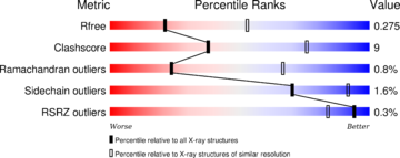
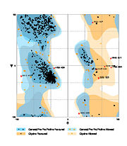
| - | The structure have been [https://www.ncbi.nlm.nih.gov/pubmed/24018048 published in the year 2013 by Qilu Ye et al.] | + | The structure have been [https://www.ncbi.nlm.nih.gov/pubmed/24018048 published in the year 2013 by Qilu Ye et al.] <ref>PMID:24018048</ref> in [http://www.sciencedirect.com/science/journal/08986568 ''Cellular Signaling'']. For their experiments they used [https://en.wikipedia.org/wiki/X-ray_crystallography#X-ray_diffraction x-ray diffraction]. The [http://www.wwpdb.org/validation/2016/XrayValidationReportHelp PDB validation] obtained a Resolutionof 3.0 Å, a free R-value of 0.273 and a work R-value of 0.241 [http://www.rcsb.org/pdb/explore/explore.do?structureId=4IL1]. |
<br style="clear:both" /> | <br style="clear:both" /> | ||
| Line 43: | Line 43: | ||
== Principle of action == | == Principle of action == | ||
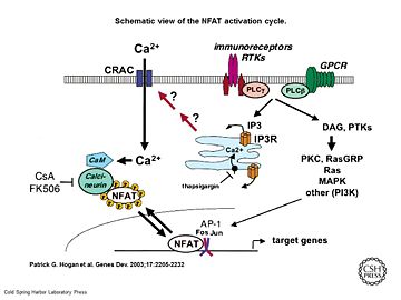
| - | [[Image:Genes Dev. 2003 Sep 17(18) 2205-32, Figure 1.jpg|thumb|upright=2|Schematic view of the NFAT activation cycle | + | [[Image:Genes Dev. 2003 Sep 17(18) 2205-32, Figure 1.jpg|thumb|upright=2|Schematic view of the NFAT activation cycle <ref>PMID:12975316</ref>]] |
As a response of receptor tyrosine kinase [https://en.wikipedia.org/wiki/Receptor_tyrosine_kinase(RTK)] activation as well as G protein-coupled receptor [https://en.wikipedia.org/wiki/G_protein%E2%80%93coupled_receptor (GCPR)] activation the [[Phospholipase C]] [https://en.wikipedia.org/wiki/Phospholipase_C (PLC)] catalyse the hydrolysis of [https://en.wikipedia.org/wiki/Phosphatidylinositol_4,5-bisphosphate PIP2] to [https://en.wikipedia.org/wiki/Inositol_trisphosphate IP3] and [https://en.wikipedia.org/wiki/Diglyceride DAG]. IP3 activates the [[Inositol 1,4,5-Trisphosphate Receptor]] and therby leads to an increasing amount of the second Messenger Ca2+ in the cytoplasma. | As a response of receptor tyrosine kinase [https://en.wikipedia.org/wiki/Receptor_tyrosine_kinase(RTK)] activation as well as G protein-coupled receptor [https://en.wikipedia.org/wiki/G_protein%E2%80%93coupled_receptor (GCPR)] activation the [[Phospholipase C]] [https://en.wikipedia.org/wiki/Phospholipase_C (PLC)] catalyse the hydrolysis of [https://en.wikipedia.org/wiki/Phosphatidylinositol_4,5-bisphosphate PIP2] to [https://en.wikipedia.org/wiki/Inositol_trisphosphate IP3] and [https://en.wikipedia.org/wiki/Diglyceride DAG]. IP3 activates the [[Inositol 1,4,5-Trisphosphate Receptor]] and therby leads to an increasing amount of the second Messenger Ca2+ in the cytoplasma. | ||
Calcineurin is activated by [http://www.ebi.ac.uk/interpro/potm/2003_3/Page_1.htm Calmodulin], a calcium-binding protein. Calmodulin interacts with the calmodulin-binding/regulatory region of Calcineurin. That binding leads to a conformational change in the autoinhibitory domain and remove it from the active site (doi:10.1016/j.jmb.2011.11.008). | Calcineurin is activated by [http://www.ebi.ac.uk/interpro/potm/2003_3/Page_1.htm Calmodulin], a calcium-binding protein. Calmodulin interacts with the calmodulin-binding/regulatory region of Calcineurin. That binding leads to a conformational change in the autoinhibitory domain and remove it from the active site (doi:10.1016/j.jmb.2011.11.008). | ||
| - | It has been reported that Calcineurin activates the transcription factor [https://de.wikipedia.org/wiki/NF-AT NFAT] by forming a complex and dephosporylation | + | It has been reported that Calcineurin activates the transcription factor [https://de.wikipedia.org/wiki/NF-AT NFAT] by forming a complex and dephosporylation <ref>PMID:17502104</ref>. Following, the factor enters the nucleus and activates the expression of Interleukin-2. |
<br style="clear:both" /> | <br style="clear:both" /> | ||
| Line 54: | Line 54: | ||
== Binding Partners == | == Binding Partners == | ||
The main partners of interaction are [https://en.wikipedia.org/wiki/Calmodulin Calmodulin],NFATc1, NFATc2 and NFATc3. | The main partners of interaction are [https://en.wikipedia.org/wiki/Calmodulin Calmodulin],NFATc1, NFATc2 and NFATc3. | ||
| - | Many of the calcineurin substrates’ contain a PxIxIT motif. Among them, beside the phosphorylated forms of NFAT we can also mentioned; cAMP response element binding protein (CREB), PP1, microtubule-associated protein tau and glycogen synthase kinase-3 beta (GSK- 3) | + | Many of the calcineurin substrates’ contain a PxIxIT motif. Among them, beside the phosphorylated forms of NFAT we can also mentioned; cAMP response element binding protein (CREB), PP1, microtubule-associated protein tau and glycogen synthase kinase-3 beta (GSK- 3)<ref>PMID: 17666045</ref><ref>PMID: 22676853</ref><ref>PMID:14701880</ref><ref>PMID: 7515479</ref>. |
| - | Calcineurin is inhibited by the immunosuppressive drugs tacrolismus (FK506) or cyclosporine A (CsA). CsA and FK506 conduct their therapeutic role thought binding to the [https://en.wikipedia.org/wiki/Immunophilins immunophilins] cyclophilin and FK506 binding protein (FK506BP) respectively. The complexes CsA-cyclophilin and FK506-FK506BP bind then to calcineurin in a calcium-dependent manner thus inhibiting its phosphatase activity. Therefore the addition of these drugs to lymphocytes T prevent NFAT translocation to the nucleus and the subsequent activation its target gene.That's why FK506 and CsA are use in the treatment of various immune-mediated diseases. However since calcineurin is is widely expressed in non-haemopoietic tissues like the kidney and the hearth, both drugs present a long term toxicity and can lead to deleterious effect to these Organs | + | Calcineurin is inhibited by the immunosuppressive drugs tacrolismus (FK506) or cyclosporine A (CsA). CsA and FK506 conduct their therapeutic role thought binding to the [https://en.wikipedia.org/wiki/Immunophilins immunophilins] cyclophilin and FK506 binding protein (FK506BP) respectively. The complexes CsA-cyclophilin and FK506-FK506BP bind then to calcineurin in a calcium-dependent manner thus inhibiting its phosphatase activity. Therefore the addition of these drugs to lymphocytes T prevent NFAT translocation to the nucleus and the subsequent activation its target gene.That's why FK506 and CsA are use in the treatment of various immune-mediated diseases. However since calcineurin is is widely expressed in non-haemopoietic tissues like the kidney and the hearth, both drugs present a long term toxicity and can lead to deleterious effect to these Organs <ref>PMID: 8811062</ref>, (http://www.uptodate.com/contents/pharmacology-of-cyclosporine-and-tacrolimus). |
'''Cofactors''': | '''Cofactors''': | ||
| - | Calcineurin belong to the family of [https://en.wikipedia.org/wiki/Metalloprotein metalloprotein]. To conduct its activity it requires the presence of Fe3+ and Zn2+ ions in the active site (one per subunit).Superoxide dismutase has been shown to protect calcineurin from inactivation by preventing Fe3+ from oxidation. Thus after activation of calcineurin by calmodulin, the AID is displaced from the catalytic core exposing Fe3+ to oxidation | + | Calcineurin belong to the family of [https://en.wikipedia.org/wiki/Metalloprotein metalloprotein]. To conduct its activity it requires the presence of Fe3+ and Zn2+ ions in the active site (one per subunit).Superoxide dismutase has been shown to protect calcineurin from inactivation by preventing Fe3+ from oxidation. Thus after activation of calcineurin by calmodulin, the AID is displaced from the catalytic core exposing Fe3+ to oxidation <ref>PMID: 8837775</ref>(Calmodulin and Signal Transduction (p184), Linda J. Van Eldik,D. Martin Watterson (1998)). |
| Line 68: | Line 68: | ||
== Related health defects == | == Related health defects == | ||
| - | Calcineurin hyperactivation thought dysregulation of the Ca2+ dynamic have been show to play a critical role in several diseases like Rheumatoid arthritis (RA), Schizophrenia ,Diabetes, Systemic Lupus Erythematosus as well as Alzheimer diseases (http://www.uptodate.com/contents/pharmacology-of-cyclosporine-and-tacrolimus) | + | Calcineurin hyperactivation thought dysregulation of the Ca2+ dynamic have been show to play a critical role in several diseases like Rheumatoid arthritis (RA), Schizophrenia ,Diabetes, Systemic Lupus Erythematosus as well as Alzheimer diseases (http://www.uptodate.com/contents/pharmacology-of-cyclosporine-and-tacrolimus)<ref>PMID: 12851457</ref><ref>PMID: 16988714</ref><ref>PMID:20421909</ref><ref>PMID: 22654726</ref>.In order to fight those health defects calmodulin inhibitors can be administrated. |
== Human/Rat calcineurin comparison == | == Human/Rat calcineurin comparison == | ||
Revision as of 11:17, 15 January 2017
Structure Rat Calcineurin
| |||||||||||


