We apologize for Proteopedia being slow to respond. For the past two years, a new implementation of Proteopedia has been being built. Soon, it will replace this 18-year old system. All existing content will be moved to the new system at a date that will be announced here.
User:Estelle Metzger/Sandbox
From Proteopedia
(Difference between revisions)
| Line 12: | Line 12: | ||
[[Image:Roco4.jpg|thumb| Linear structure of Roco4 <ref name="Bernd2">doi: 10.3389/fnmol.2014.00032</ref>|left|upright=3]] | [[Image:Roco4.jpg|thumb| Linear structure of Roco4 <ref name="Bernd2">doi: 10.3389/fnmol.2014.00032</ref>|left|upright=3]] | ||
| + | |||
| + | |||
| + | |||
| + | |||
| + | |||
The ROC domain possesses five G motifs that are required for guanine nucleotid binding. This domain presents some similarities with the proteins of the ras family. | The ROC domain possesses five G motifs that are required for guanine nucleotid binding. This domain presents some similarities with the proteins of the ras family. | ||
| Line 18: | Line 23: | ||
The Roco4 kinase structure consists of a canonical, two-lobed kinase structure, with an adenine nucleotide bound in the conventional nucleotide-binding pocket. It contains the conserved alphaC-helix and an anti-parallel beta sheets in the smaller N-terminal lobe. Other Alpha-helices and the activation loop with the conserved N-terminal DFG motif are localized in the bigger C-terminal lobe. | The Roco4 kinase structure consists of a canonical, two-lobed kinase structure, with an adenine nucleotide bound in the conventional nucleotide-binding pocket. It contains the conserved alphaC-helix and an anti-parallel beta sheets in the smaller N-terminal lobe. Other Alpha-helices and the activation loop with the conserved N-terminal DFG motif are localized in the bigger C-terminal lobe. | ||
The activation loop and alphaC-helix together form the catalytic site of the kinase, an ATP binding site formed by a cleft between the two lobes. | The activation loop and alphaC-helix together form the catalytic site of the kinase, an ATP binding site formed by a cleft between the two lobes. | ||
| - | For catalysis, the formation of a polar contact is essential. This polar contact takes place between Roco4 Lys1055 from the beta3-strand and the Glu1078 from the alphaC-helix. The amino acids Asp makes contact with all three ATP phosphates either directly or via coordination of a magnesium ion. Moreover, the amino acid Phe makes hydrophobic contacts to the alphaC-helix and the HxD motif, and leads for the correct positioning of the DFG motif. Roco4 has two conformation, an active conformation and an inactive conformation. These conformations depend of the conformation of the DFG motif : a DFG-in (active) and a DFG-out (inactive) conformation. Therefore, in the structure of active Roco4 kinase, the activation loop is visible and ordered. In contrast, in the structure of inactive Roco4 kinase, the activation loop is not | + | For catalysis, the formation of a polar contact is essential. This polar contact takes place between Roco4 Lys1055 from the beta3-strand and the Glu1078 from the alphaC-helix. The amino acids Asp makes contact with all three ATP phosphates either directly or via coordination of a magnesium ion. Moreover, the amino acid Phe makes hydrophobic contacts to the alphaC-helix and the HxD motif, and leads for the correct positioning of the DFG motif. Roco4 has two conformation, an active conformation and an inactive conformation. These conformations depend of the conformation of the DFG motif : a DFG-in (active) and a DFG-out (inactive) conformation. Therefore, in the structure of active Roco4 kinase, the activation loop is visible and ordered. In contrast, in the structure of inactive Roco4 kinase, the activation loop is not visible. (Huse and Kuriyan, 2002 ; Taylor and Kornev, 2011). |
In most kinases, there is a mechanism to switch from an inactive to an active state. | In most kinases, there is a mechanism to switch from an inactive to an active state. | ||
This involves autophosphorylation of some residues in the activation loop. . Autophosphorylation not only results in the reorientation of the activation loop, but often also alters ATP binding and/or interaction with substrates. (Huse and Kuriyan 2002 kornev). In Roco4 kinase, there are four phosphorylation sites in the activation loop : Ser1181, Ser1184, Ser1187, and Ser1189. | This involves autophosphorylation of some residues in the activation loop. . Autophosphorylation not only results in the reorientation of the activation loop, but often also alters ATP binding and/or interaction with substrates. (Huse and Kuriyan 2002 kornev). In Roco4 kinase, there are four phosphorylation sites in the activation loop : Ser1181, Ser1184, Ser1187, and Ser1189. | ||
| - | The structure of ''Dictyostelium'' Roco4 kinase in complex with the LRRK2 inhibitor H1152 allows us to see that Roco4 and other Roco family proteins are essential for the optimization of the current, and identification of new LRRK2 kinase inhibitor. To have a Roco4 protein which have an active site resembling human LRRK2, researchers use a ''Dictyostelium'' Roco4 mutant (F1107L and F1161L) which is called | + | The structure of ''Dictyostelium'' Roco4 kinase in complex with the LRRK2 inhibitor H1152 allows us to see that Roco4 and other Roco family proteins are essential for the optimization of the current, and identification of new LRRK2 kinase inhibitor. To have a Roco4 protein which have an active site resembling human LRRK2, researchers use a ''Dictyostelium'' Roco4 mutant (F1107L and F1161L) which is called humanized Roco4.<ref name="Bernd"/> |
== LRRK2-IN-1 == | == LRRK2-IN-1 == | ||
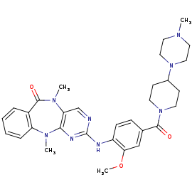
[[Image:4K4-270.png|thumb|LRRK2-IN-1 structure|upright=2]] | [[Image:4K4-270.png|thumb|LRRK2-IN-1 structure|upright=2]] | ||
| - | <scene name='75/751216/Lrrk2-in-1/1'>LRRK2-IN-1</scene> | + | <scene name='75/751216/Lrrk2-in-1/1'>LRRK2-IN-1</scene> is the first identified LRRK2-specific inhibitor, which is now a common tool compound for the LRRK2 research community. LRRK2-IN-1 has a 2-amino-5,11- dimethyl-5H-benzo[e]pyrimido[5,4-b][1,4]diazepine-6(11H)-one scaffold. |
The function is of LRRK2-In-1 is to dephosphorylate LRRK2 residues Ser910 and Ser935 in the kidney, but not in the brain. This compound is not capable of crossing the blood-brain barrier. | The function is of LRRK2-In-1 is to dephosphorylate LRRK2 residues Ser910 and Ser935 in the kidney, but not in the brain. This compound is not capable of crossing the blood-brain barrier. | ||
The structure of LRRK2-In-1 does not stabilize the active conformation. Indeed, the activation loop is poorly resolved indicating that it is flexible. Moreover, it presents a closure of the glycine-rich loop in the inhibitor structure.<ref name="Bernd"/> | The structure of LRRK2-In-1 does not stabilize the active conformation. Indeed, the activation loop is poorly resolved indicating that it is flexible. Moreover, it presents a closure of the glycine-rich loop in the inhibitor structure.<ref name="Bernd"/> | ||
Revision as of 17:17, 26 January 2017
Humanized Roco4 bound to LRRK2-IN-1
| |||||||||||
References
- ↑ 1.0 1.1 1.2 1.3 1.4 1.5 1.6 1.7 Gilsbach BK, Messias AC, Ito G, Sattler M, Alessi DR, Wittinghofer A, Kortholt A. Structural Characterization of LRRK2 Inhibitors. J Med Chem. 2015 May 1. PMID:25897865 doi:http://dx.doi.org/10.1021/jm5018779
- ↑ Gilsbach BK, Kortholt A. Structural biology of the LRRK2 GTPase and kinase domains: implications for regulation. Front Mol Neurosci. 2014 May 5;7:32. doi: 10.3389/fnmol.2014.00032. eCollection, 2014. PMID:24847205 doi:http://dx.doi.org/10.3389/fnmol.2014.00032


