User:Ophelie Lefort/Sandbox
From Proteopedia
(Difference between revisions)
| Line 11: | Line 11: | ||
== Structural highlights == | == Structural highlights == | ||
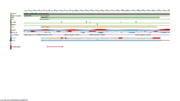
| - | + | The protein has a 283 amino-acids long sequence with two mainly domains [[Image:Linear structure of PRSS57.png | thumb]]. It forms a kind of elongate sphere of approximately 70Å large and 150Å long (dimensions: 70,37Å X 70,37Å X 105,02Å). The protein is composed of signal peptide (1-31) which leads to the location in azurophil granules [http://www.jimmunol.org/content/191/5/2700] and allows its excretion. In addition there is a protease domain (32-283) which is a trypsin-like domain with a trypsin-like <scene name='75/751133/Active_site/1'>active site</scene> , according to the specificity for P1-Arg residues, but this domain can be an elastase-like active site according to the primary sequence (because of the presence of a swallow S1 pocket) specific to small aliphatic residues.<ref>S.. Jack Lin, Ken C. Dong, Charles Eigenbrot, Menno van Lookeren Campagne, Daniel Kirchhofer Structures of Neutrophil Serine Protease 4 Reveal an Unusual Mechanism of Substrate Recognition by a Trypsin-Fold Protease DOI: http://dx.doi.org/10.1016/j.str.2014.07.008</ref> | |
| - | + | The <scene name='75/751133/Active_site/1'>active site</scene> is form by 4 amino acids: Gly(189), Phe(190), Ser(216), D(226) | |
| - | The protein has a 283 amino-acids long sequence with two mainly domains. It forms a kind of elongate sphere of approximately 70Å large and 150Å long (dimensions: 70,37Å X 70,37Å X 105,02Å). The protein is composed of signal peptide (1-31) which leads to the location in azurophil granules [http://www.jimmunol.org/content/191/5/2700] and allows its excretion. In addition there is a protease domain (32-283) which is a trypsin-like domain with a trypsin-like <scene name='75/751133/Active_site/1'>active site</scene> , according to the specificity for P1-Arg residues, but this domain can be an elastase-like active site according to the primary sequence (because of the presence of a swallow S1 pocket) specific to small aliphatic residues.<ref>S.. Jack Lin, Ken C. Dong, Charles Eigenbrot, Menno van Lookeren Campagne, Daniel Kirchhofer Structures of Neutrophil Serine Protease 4 Reveal an Unusual Mechanism of Substrate Recognition by a Trypsin-Fold Protease DOI: http://dx.doi.org/10.1016/j.str.2014.07.008</ref> | + | |
| - | The <scene name='75/751133/Active_site/1'>active site</scene> is | + | |
The residue F190 obstructs the active site which could normally not links a P1-Arg. However, a study <ref>Natascha C. Perera, Karl-Heinz Wiesmüller, Maria Torp Larsen, Beate Schacher, Peter Eickholz, Niels Borregaard and Dieter E. Jenne NSP4 Is Stored in Azurophil Granules and Released by Activated Neutrophils as Active Endoprotease with Restricted Specificity DOI: https://doi.org/10.4049/jimmunol.1301293</ref> considered the possibility that the two residues S216 and F190 of the active site can form a flexible gate which allows P1-Arg to enter. Then, the link between the active site and P1-Arg can be stabilized by a salt bridge interaction between S1-D226 and P1-Arg. | The residue F190 obstructs the active site which could normally not links a P1-Arg. However, a study <ref>Natascha C. Perera, Karl-Heinz Wiesmüller, Maria Torp Larsen, Beate Schacher, Peter Eickholz, Niels Borregaard and Dieter E. Jenne NSP4 Is Stored in Azurophil Granules and Released by Activated Neutrophils as Active Endoprotease with Restricted Specificity DOI: https://doi.org/10.4049/jimmunol.1301293</ref> considered the possibility that the two residues S216 and F190 of the active site can form a flexible gate which allows P1-Arg to enter. Then, the link between the active site and P1-Arg can be stabilized by a salt bridge interaction between S1-D226 and P1-Arg. | ||
The hypothesis of the flexible gate was confirmed by the same study. The mutations of S216 only, F190 only or both together show a forced full open gate is more efficient than a forced partially open gate. | The hypothesis of the flexible gate was confirmed by the same study. The mutations of S216 only, F190 only or both together show a forced full open gate is more efficient than a forced partially open gate. | ||
| - | The conclusion is that the protease domain is a trypsin-like domain.[[Image:Active site | + | The conclusion is that the protease domain is a trypsin-like domain.[[Image:Active site.jpg | thumb]] |
| Line 23: | Line 21: | ||
Like in other trypsin-like proteases <scene name='75/751133/F190/1'>D226</scene> is inaccessible to the substrate but helps stabilize the closed S1 pocket by forming an H-bond with the <scene name='75/751133/F190/1'>F190</scene> amide. | Like in other trypsin-like proteases <scene name='75/751133/F190/1'>D226</scene> is inaccessible to the substrate but helps stabilize the closed S1 pocket by forming an H-bond with the <scene name='75/751133/F190/1'>F190</scene> amide. | ||
| - | NSP4 compared to the other trypsin-like proteases has some specificity as the arginine side chain movement from the canonical "down" to the noncanonical "up" position in NSP4, which is accomplished by a rotation of the Chi2 angle by 160°, this is possible thanks to <scene name='75/751133/F190/1'>F190</scene> which provides a hydrophobic platform that interacts with the aliphatic portion of the P1-arginine side chain. All other residue positions are preserved. The specificity for P1-arginine is conferred by H-bonds between guanidinium group and <scene name='75/751133/H_bounds/1'>three H-bonds acceptors (S216, S192, G217)</scene>. This specificity allows NSP4 to cleave | + | NSP4 compared to the other trypsin-like proteases has some specificity as the arginine side chain movement from the canonical "down" to the noncanonical "up" position in NSP4, which is accomplished by a rotation of the Chi2 angle by 160°, this is possible thanks to <scene name='75/751133/F190/1'>F190</scene> which provides a hydrophobic platform that interacts with the aliphatic portion of the P1-arginine side chain. All other residue positions are preserved. The specificity for P1-arginine is conferred by H-bonds between guanidinium group and <scene name='75/751133/H_bounds/1'>three H-bonds acceptors (S216, S192, G217)</scene>. This specificity allows NSP4 to cleave citrulline, which is not cleaved by other trypsin-like proteases because of its non-charged propriety, and the impossibility to do salt-bridges. Other trypsin-like proteases are not able to cleave methylarginine neither because of a steric clash with the methyl group, which is not an issue for NSP4 since the methyl group is exposed to the solvent. The capacity to cleave these post-translationally modified arginine utility may be to act against microbial and virulence factors containing modified arginine or to interact with chemokines. <ref>Lin, S. Jack, Ken C. Dong, Charles Eigenbrot, Menno van Lookeren Campagne, and Daniel Kirchhofer. “Structures of Neutrophil Serine Protease 4 Reveal an Unusual Mechanism of Substrate Recognition by a Trypsin-Fold Protease.” Structure 22, no. 9 (September 2, 2014): 1333–40.[http://dx.doi.org/10.1016/j.str.2014.07.008]</ref> |
| - | <scene name='75/751133/Signal_peptide/1'>N terminus of NSP4</scene> has been shown to be cleaved off by | + | <scene name='75/751133/Signal_peptide/1'>N terminus of NSP4</scene> has been shown to be cleaved off by Cathepsin C and occurs during the translocation into the endoplasmic reticulum before the enzyme is stored in cytoplasmic granules. Natural serine protease inhibitors of NSP4 are present in human plasma and can form covalent NSP4-serpin complexes in vitro (antithrombin for example which act as a suicide substrate with NSP4). However, in vivo, antithrombin can not trap NSP4 because of the presence of other neutrophil proteases.<ref>Perera, Natascha C., Karl-Heinz Wiesmüller, Maria Torp Larsen, Beate Schacher, Peter Eickholz, Niels Borregaard, and Dieter E. Jenne. “NSP4 Is Stored in Azurophil Granules and Released by Activated Neutrophils as Active Endoprotease with Restricted Specificity.” The Journal of Immunology 191, no. 5 (September 1, 2013) [https://doi.org/10.4049/jimmunol.1301293]</ref> |
Revision as of 10:51, 27 January 2017
Neutrophil Serine 4 or Serine Protease 57 (4Q7X)
| |||||||||||
References
[3] NSP4 is stored in azurophil granules and released by activated neutrophils as active endoprotease with restricted specificity, NCBI
[4] NSP4, an elastase-related protease in human neutrophils with arginine specificity, NCBI
[5] Tailor-made inflammation: how neutrophil serine proteases modulate the inflammatory response, NCBI
[6] Role of neutrophils in innate immunity: a systems biology-level approach, NCBI
[7] Neutrophils: Their Role in Innate and Adaptive Immunity, NCBI
[8] Neutrophil extracellular traps kill bacteria, NCBI
[9] Tailor-made inflammation: how neutrophil serine proteases modulate the inflammatory response, NCBI
- ↑ S.. Jack Lin, Ken C. Dong, Charles Eigenbrot, Menno van Lookeren Campagne, Daniel Kirchhofer Structures of Neutrophil Serine Protease 4 Reveal an Unusual Mechanism of Substrate Recognition by a Trypsin-Fold Protease DOI: http://dx.doi.org/10.1016/j.str.2014.07.008
- ↑ Natascha C. Perera, Karl-Heinz Wiesmüller, Maria Torp Larsen, Beate Schacher, Peter Eickholz, Niels Borregaard and Dieter E. Jenne NSP4 Is Stored in Azurophil Granules and Released by Activated Neutrophils as Active Endoprotease with Restricted Specificity DOI: https://doi.org/10.4049/jimmunol.1301293
- ↑ Lin, S. Jack, Ken C. Dong, Charles Eigenbrot, Menno van Lookeren Campagne, and Daniel Kirchhofer. “Structures of Neutrophil Serine Protease 4 Reveal an Unusual Mechanism of Substrate Recognition by a Trypsin-Fold Protease.” Structure 22, no. 9 (September 2, 2014): 1333–40.[1]
- ↑ Perera, Natascha C., Karl-Heinz Wiesmüller, Maria Torp Larsen, Beate Schacher, Peter Eickholz, Niels Borregaard, and Dieter E. Jenne. “NSP4 Is Stored in Azurophil Granules and Released by Activated Neutrophils as Active Endoprotease with Restricted Specificity.” The Journal of Immunology 191, no. 5 (September 1, 2013) [2]


