This old version of Proteopedia is provided for student assignments while the new version is undergoing repairs. Content and edits done in this old version of Proteopedia after March 1, 2026 will eventually be lost when it is retired in about June of 2026.
Apply for new accounts at the new Proteopedia. Your logins will work in both the old and new versions.
Peptide N-glycanase
From Proteopedia
(Difference between revisions)
| Line 11: | Line 11: | ||
PNG F is used in studying of glycoproteins. | PNG F is used in studying of glycoproteins. | ||
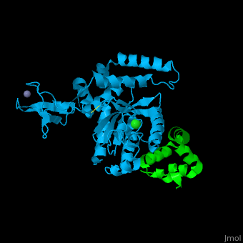
== Structural highlights == | == Structural highlights == | ||
| + | *<scene name='68/684794/Cv/2'>Zn coordination site contains 4 cysteins</scene>. | ||
| + | *<scene name='68/684794/Cv/3'>Cl coordination site</scene>. | ||
</StructureSection> | </StructureSection> | ||
== 3D Structures of PNGase == | == 3D Structures of PNGase == | ||
Revision as of 09:48, 28 August 2017
| |||||||||||
3D Structures of PNGase
Updated on 28-August-2017
References
- ↑ Tanabe K, Lennarz WJ, Suzuki T. A cytoplasmic peptide: N-glycanase. Methods Enzymol. 2006;415:46-55. PMID:17116467 doi:http://dx.doi.org/10.1016/S0076-6879(06)15004-1
- ↑ Norris GE, Stillman TJ, Anderson BF, Baker EN. The three-dimensional structure of PNGase F, a glycosylasparaginase from Flavobacterium meningosepticum. Structure. 1994 Nov 15;2(11):1049-59. PMID:7881905
- ↑ Suzuki T. The cytoplasmic peptide:N-glycanase (Ngly1)-basic science encounters a human genetic disorder. J Biochem. 2015 Jan;157(1):23-34. doi: 10.1093/jb/mvu068. Epub 2014 Nov 13. PMID:25398991 doi:http://dx.doi.org/10.1093/jb/mvu068
