This old version of Proteopedia is provided for student assignments while the new version is undergoing repairs. Content and edits done in this old version of Proteopedia after March 1, 2026 will eventually be lost when it is retired in about June of 2026.
Apply for new accounts at the new Proteopedia. Your logins will work in both the old and new versions.
Ephrin
From Proteopedia
(Difference between revisions)
| Line 1: | Line 1: | ||
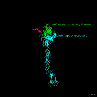
| - | <StructureSection load=' | + | <StructureSection load='' size='340' side='right' caption='Structure of glycosylated human ephrin-A5 receptor-binding domain (green) complex with ephrin type A receptor 2 (cyan) (PDB code [[3mx0]]).' scene='59/594659/Cv/1'> |
== Function == | == Function == | ||
Revision as of 10:30, 20 October 2017
| |||||||||||
3D structures of ephrin
Updated on 20-October-2017
References
- ↑ Egea J, Klein R. Bidirectional Eph-ephrin signaling during axon guidance. Trends Cell Biol. 2007 May;17(5):230-8. Epub 2007 Apr 8. PMID:17420126 doi:http://dx.doi.org/10.1016/j.tcb.2007.03.004
- ↑ Himanen JP, Yermekbayeva L, Janes PW, Walker JR, Xu K, Atapattu L, Rajashankar KR, Mensinga A, Lackmann M, Nikolov DB, Dhe-Paganon S. Architecture of Eph receptor clusters. Proc Natl Acad Sci U S A. 2010 May 26. PMID:20505120
