Eric Comeau/ sandbox
From Proteopedia
(Difference between revisions)
| Line 5: | Line 5: | ||
== Physical model == | == Physical model == | ||
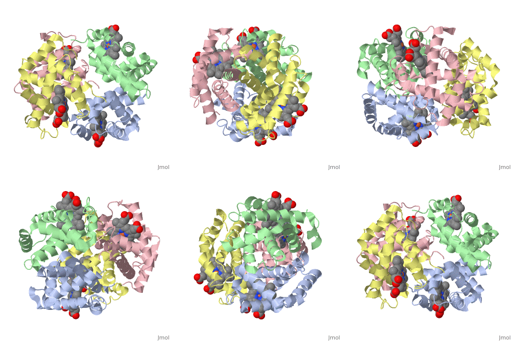
| - | The 3D-printed models are of the tetramer, with one beta subunit valine marked with a blue rod. One hydrophobic surface patch is marked with a hole. In the browser, the <scene name='61/611454/One_with_val/1'>tetramer</scene> is shown in red (alpha subunits) and orangered (beta subunits), with both valines shown in blue. In a second scene, <scene name='61/611454/Two_with_val/1'>two interacting tetramers</scene> are shown. You can see that one of the four valines is buried in the contact area | + | The 3D-printed models are of the tetramer, with one beta subunit valine marked with a blue rod. One hydrophobic surface patch is marked with a hole. In the browser, the <scene name='61/611454/One_with_val/1'>tetramer</scene> is shown in red (alpha subunits) and orangered (beta subunits), with both valines shown in blue. In a second scene, <scene name='61/611454/Two_with_val/1'>two interacting tetramers</scene> are shown. You can see that one of the four valines is buried in the contact area between the two tetramers. The <scene name='61/611454/Val_and_patch/2'>hydrophobic patch</scene> (shown in gray) consists of Phe 85 and Leu 88 side chains in an indentation of the protein's surface. |
== Sickle Cell Anemia == | == Sickle Cell Anemia == | ||
Revision as of 19:56, 11 January 2018
Sickle Cell Hemoglobin
| |||||||||||
References
- ↑ Hanson, R. M., Prilusky, J., Renjian, Z., Nakane, T. and Sussman, J. L. (2013), JSmol and the Next-Generation Web-Based Representation of 3D Molecular Structure as Applied to Proteopedia. Isr. J. Chem., 53:207-216. doi:http://dx.doi.org/10.1002/ijch.201300024
- ↑ Herraez A. Biomolecules in the computer: Jmol to the rescue. Biochem Mol Biol Educ. 2006 Jul;34(4):255-61. doi: 10.1002/bmb.2006.494034042644. PMID:21638687 doi:10.1002/bmb.2006.494034042644

