We apologize for Proteopedia being slow to respond. For the past two years, a new implementation of Proteopedia has been being built. Soon, it will replace this 18-year old system. All existing content will be moved to the new system at a date that will be announced here.
Major vault protein
From Proteopedia
(Difference between revisions)
| Line 15: | Line 15: | ||
MVP was found to enhance the expression of the anti-apoptotic protein [[bcl-2]] in senescent human fibroblasts <ref> Ryu, S. J., An, H. J., Oh, Y. S., Choi, H. R., Ha, M. K., and Park, S. C. (2008) On the role of major vault protein in the resistance of senescent human diploid fibroblasts to apoptosis. Cell Death Differ. doi: 10.1038/cdd.2008.96.</ref>. By binding to [[COP1]], which is an [[E3 ligase]], MVP forms an interaction which is essential for the degradation of [[c-June]]. This degradation is important in senescent human fibroblasts regarding the modulation of the anti-apoptotic protein bcl-2, and it is reduced when MVP is subjected to UV light which causes it to be tyrosine-phosphorylated. | MVP was found to enhance the expression of the anti-apoptotic protein [[bcl-2]] in senescent human fibroblasts <ref> Ryu, S. J., An, H. J., Oh, Y. S., Choi, H. R., Ha, M. K., and Park, S. C. (2008) On the role of major vault protein in the resistance of senescent human diploid fibroblasts to apoptosis. Cell Death Differ. doi: 10.1038/cdd.2008.96.</ref>. By binding to [[COP1]], which is an [[E3 ligase]], MVP forms an interaction which is essential for the degradation of [[c-June]]. This degradation is important in senescent human fibroblasts regarding the modulation of the anti-apoptotic protein bcl-2, and it is reduced when MVP is subjected to UV light which causes it to be tyrosine-phosphorylated. | ||
===MVP and vaults in signal regulation and transport platforms=== | ===MVP and vaults in signal regulation and transport platforms=== | ||
| - | Though the inner cavity of the vault particle created by MVP was reported to accommodate an unknown inner mass <ref> Kong, L. B., Siva, A. C., Rome, L. H., and Stewart, P. L. (1999) | + | Though the inner cavity of the vault particle created by MVP was reported to accommodate an unknown inner mass <ref name=kong> Kong, L. B., Siva, A. C., Rome, L. H., and Stewart, P. L. (1999) |
Structure of the vault, a ubiquitous celular component. | Structure of the vault, a ubiquitous celular component. | ||
Structure Fold Des. 7, 371 – 379.</ref>, and though vaults have known qualities like rapid movement to [[lipid raft]]s, unique subcellular localization <ref> Slesina, M., Inman, E. M., Rome, L. H., and Volknandt, W. | Structure Fold Des. 7, 371 – 379.</ref>, and though vaults have known qualities like rapid movement to [[lipid raft]]s, unique subcellular localization <ref> Slesina, M., Inman, E. M., Rome, L. H., and Volknandt, W. | ||
| Line 41: | Line 41: | ||
constitutively photomorphogenic 1, negatively regulates cJun-mediated | constitutively photomorphogenic 1, negatively regulates cJun-mediated | ||
activator protein 1 transcription in mammalian | activator protein 1 transcription in mammalian | ||
| - | cells. Cancer Res. 65, 5835 – 5840.</ref>, thus allowing it to bind to the MAPK [[Erk]] and the tyrosine phosphorylase [[SHP-2]]<ref> Kolli, S., Zito, C. I., Mossink, M. H., Wiemer, E. A., and | + | cells. Cancer Res. 65, 5835 – 5840.</ref>, thus allowing it to bind to the MAPK [[Erk]] and the tyrosine phosphorylase [[SHP-2]]<ref name=kolli> Kolli, S., Zito, C. I., Mossink, M. H., Wiemer, E. A., and |
Bennett, A. M. (2004) The major vault protein is a novel | Bennett, A. M. (2004) The major vault protein is a novel | ||
substrate for the tyrosine phosphatase SHP-2 and scaffold | substrate for the tyrosine phosphatase SHP-2 and scaffold | ||
| Line 48: | Line 48: | ||
Ryu, S. H., and Suh, P. G. (2006) Crosstalk between Src and | Ryu, S. H., and Suh, P. G. (2006) Crosstalk between Src and | ||
major vault protein in epidermal growth factor-dependent cell | major vault protein in epidermal growth factor-dependent cell | ||
| - | signalling. Febs J. 273, 793 – 804.</ref>. This data is thought to indicate that MVP might have a scaffolding function for signal transduction<ref | + | signalling. Febs J. 273, 793 – 804.</ref>. This data is thought to indicate that MVP might have a scaffolding function for signal transduction.<ref name=kolli /> |
| - | + | ||
| - | + | ||
| - | + | ||
| - | + | ||
* MVP, together with the vRNA of vaults, were found to bind to [[Estrogen]] receptors by interacting through several proto-NLS found on the receptors and which are in charge of the hormone-independent nuclear import [149]. | * MVP, together with the vRNA of vaults, were found to bind to [[Estrogen]] receptors by interacting through several proto-NLS found on the receptors and which are in charge of the hormone-independent nuclear import [149]. | ||
* MVP (-/-) mice are extremely prone to pseudomonas aeruginosa infections, thus it is speculated that MVP is involved in the signal transduction activating the innate-immune system to some extent[115]. | * MVP (-/-) mice are extremely prone to pseudomonas aeruginosa infections, thus it is speculated that MVP is involved in the signal transduction activating the innate-immune system to some extent[115]. | ||
== Structural highlights == | == Structural highlights == | ||
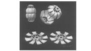
| - | MVP is highly conserved in evolution and can create the entire outer shell of the vault barrel structure, which is comprised of two identical halves. The outer shell is a closed, smooth surface without any large gaps or windows. When considering the individual MVP within a vault particle, their <scene name='78/783129/N-terminus/1'>N-terminus ( residues 113–620)</scene> forms the waist of the particle while their <scene name='78/783129/C-terminus/2'>C-terminus (residues 621-893)</scene> builds the cap and the cap/barrel junction[26]. This leads to the current belief that the N-terminus accounts for the non-covalent interactions between the identical particle halves | + | MVP is highly conserved in evolution and can create the entire outer shell of the vault barrel structure, which is comprised of two identical halves. The outer shell is a closed, smooth surface without any large gaps or windows. When considering the individual MVP within a vault particle, their <scene name='78/783129/N-terminus/1'>N-terminus ( residues 113–620)</scene> forms the waist of the particle while their <scene name='78/783129/C-terminus/2'>C-terminus (residues 621-893)</scene> builds the cap and the cap/barrel junction[26]. This leads to the current belief that the N-terminus accounts for the non-covalent interactions between the identical particle halves <ref name=Mikyas> Mikyas, Y., Makabi, M., Raval-Fernandes, S., Harrington, L., Kickhoefer, V. A., Rome, L. H., and Stewart, P. L. (2004) Cryoelectron microscopy imaging of recombinant and tissue derived vaults: localization of the MVP N termini and VPARP. J. Mol. Biol. 344, 91 – 105. </ref>. In addition, the individual MVP represents a unique protein that does not share a homology with other proteins, yet exhibits a high degree of conservation <ref name=kong /> <ref name=Mikyas /> <ref name= kick> Kickhoefer, V. A., Vasu, S. K., and Rome, L. H. (1996) Vaults |
| - | There are several domains within MVP, among the most important is the highly conserved<scene name='78/783129/C-terminus/2'> α- helical domain</scene> near the C-terminus that functions as a coiled coil which mediates an interaction between different MVPs and subsequently vault formation. The N-terminal of MVP was reported to bind Ca2+, but while it has been speculated that MVP contains at least two Ca2+-binding [[EF hand]]s in<scene name='78/783129/Ef-hand_location/1'> positions 131–143</scene> | + | are the answer, what is the question? Trends Cell Biol. 6, 174 – 178.</ref> <ref name=anderson> Anderson, D. H., Kickhoefer, V. A., Sievers, S. A., Rome, L. H., and Eisenberg, D. (2007) Draft crystal structure of the vault shell at 9-A resolution. PLoS Biol. 5, e318. </ref> <ref name=kedersha> Kedersha, N. L., and Rome, L. H. (1990) Vaults: large |
| + | cytoplasmic RNP�s that associate with cytoskeletal elements. Mol. Biol. Rep. 14, 121 – 122. </ref>- around 90% within mammals <ref name=kedersha14> Kedersha, N. L., Miquel, M. C., Bittner, D., and Rome, L. H. (1990) Vaults. II. Ribonucleoprotein structures are highly conserved among higher and lower eukaryotes. J. Cell Biol. 110, 895 – 901. </ref> <ref name=mossink 16> Mossink, M. H., van Zon, A., Scheper, R. J.,Sonneveld, P., Wiemer, E. A., Schoester, M., Houtsmuller, A. B., Scheffer, G. L., Franzel-Luiten, E., Kickhoefer, V. A., Mossink, M., Poderycki, M. J., Chan, E. K., and Rome, L. H. (2003) Vaults: a ribonucleoprotein particle involved in drug resistance? Oncogene 22, 7458 – 7467.</ref> | ||
| + | There are several domains within MVP, among the most important is the highly conserved<scene name='78/783129/C-terminus/2'> α- helical domain</scene> near the C-terminus that functions as a coiled coil which mediates an interaction between different MVPs and subsequently vault formation. The N-terminal of MVP was reported to bind Ca2+, but while it has been speculated that MVP contains at least two Ca2+-binding [[EF hand]]s in<scene name='78/783129/Ef-hand_location/1'> positions 131–143</scene> <ref name=yu 28> Yu, Z., Fotouhi-Ardakani, N., Wu, L., Maoui, M., Wang, S., Banville, D., and Shen, S. H. (2002) PTEN associates with the vault particles in HeLa cells. J. Biol. Chem. 277, 40247 – 40252. </ref> , substructure determinations by NMR could not confirm these EF hands and thus an alternative Ca2+ mechanism was suggested which included coordination by large number of <scene name='78/783129/Beta_loops/1'>acidic residues in the long β1/β2 and β2/β3 loops</scene> of multiple MVP domains <ref name=kozlov 10> Kozlov, G., Vavelyuk, O., Minailiuc, O., Banville, D., Gehring, K., and Ekiel, I. (2006) Solution structure of a two-repeat fragment of major vault protein. J. Mol. Biol. 356, 444 – 452 </ref> , in a way similar to that found in[[ integrin]]s. | ||
==The MVP gene, transcription, translation and post translation modifications== | ==The MVP gene, transcription, translation and post translation modifications== | ||
Revision as of 18:05, 16 March 2018
The Major Vault Protein
| |||||||||||
References
- ↑ Suprenant, K. A. (2002) Vault ribonucleoprotein particles: sarcophagi, gondolas, or safety deposit boxes? Biochemistry 41, 14447 – 14454
- ↑ Izquierdo, M. A., Scheffer, G. L., Flens, M. J., Shoemaker, R. H., Rome, L. H., and Scheper, R. J. (1996) Relationship of LRP-human major vault protein to in vitro and clinical resistance to anticancer drugs. Cytotechnology 19, 191 – 197.
- ↑ Mossink, M. H., van Zon, A., Franzel-Luiten, E., Schoester,M., Kickhoefer, V. A., Scheffer, G. L., Scheper, R. J.,Sonneveld, P., and Wiemer, E. A. (2002) Disruption of themurine major vault protein (MVP/LRP) gene does not induce hypersensitivity to cytostatics. Cancer Res. 62, 7298 – 7304.
- ↑ Ryu, S. J., An, H. J., Oh, Y. S., Choi, H. R., Ha, M. K., and Park, S. C. (2008) On the role of major vault protein in the resistance of senescent human diploid fibroblasts to apoptosis. Cell Death Differ. doi: 10.1038/cdd.2008.96.
- ↑ 5.0 5.1 Kong, L. B., Siva, A. C., Rome, L. H., and Stewart, P. L. (1999) Structure of the vault, a ubiquitous celular component. Structure Fold Des. 7, 371 – 379.
- ↑ Slesina, M., Inman, E. M., Rome, L. H., and Volknandt, W. (2005) Nuclear localization of the major vault protein in U373 cells. Cell Tissue Res. 321, 97 – 104.
- ↑ Herrmann, C., Golkaramnay, E., Inman, E., Rome, L., and Volknandt, W. (1999) Recombinant major vault protein is targeted to neuritic tips of PC12 cells. J. Cell Biol. 144, 1163 – 1172.
- ↑ Herrmann, C., Volknandt, W., Wittich, B., Kellner, R., and Zimmermann, H. (1996) The major vault protein (MVP100) is contained in cholinergic nerve terminals of electric ray electric organ. J. Biol. Chem. 271, 13908 – 13915.
- ↑ Kowalski, M. P., Dubouix-Bourandy, A., Bajmoczi, M., Golan, D. E., Zaidi, T., Coutinho-Sledge, Y. S., Gygi, M. P., Gygi, S. P., Wiemer, E. A., and Pier, G. B. (2007) Host resistance to lung infection mediated by major vault protein in epithelial cells. Science 317, 130 – 132.
- ↑ Steiner, E., Holzmann, K., Elbling, L., Micksche, M., and Berger, W. (2006) Cellular functions of vaults and their involvement in multidrug resistance. Curr. Drug Targets 7, 923 – 934.
- ↑ Chung, J. H., and Eng, C. (2005) Nuclear-cytoplasmic partitioning of phosphatase and tensin homologue deleted on chromosome 10 (PTEN) differentially regulates the cell cycle and apoptosis. Cancer Res. 65, 8096 – 8100
- ↑ Yi, C., Li, S., Chen, X., Wiemer, E. A., Wang, J., Wei, N., and Deng, X. W. (2005) Major vault protein, in concert with constitutively photomorphogenic 1, negatively regulates cJun-mediated activator protein 1 transcription in mammalian cells. Cancer Res. 65, 5835 – 5840.
- ↑ 13.0 13.1 Kolli, S., Zito, C. I., Mossink, M. H., Wiemer, E. A., and Bennett, A. M. (2004) The major vault protein is a novel substrate for the tyrosine phosphatase SHP-2 and scaffold protein in epidermal growth factor signaling. J. Biol. Chem. 279, 29374 – 29385.
- ↑ Kim, E., Lee, S., Mian, M. F., Yun, S. U., Song, M., Yi, K. S., Ryu, S. H., and Suh, P. G. (2006) Crosstalk between Src and major vault protein in epidermal growth factor-dependent cell signalling. Febs J. 273, 793 – 804.
- ↑ 15.0 15.1 Mikyas, Y., Makabi, M., Raval-Fernandes, S., Harrington, L., Kickhoefer, V. A., Rome, L. H., and Stewart, P. L. (2004) Cryoelectron microscopy imaging of recombinant and tissue derived vaults: localization of the MVP N termini and VPARP. J. Mol. Biol. 344, 91 – 105.
- ↑ Kickhoefer, V. A., Vasu, S. K., and Rome, L. H. (1996) Vaults are the answer, what is the question? Trends Cell Biol. 6, 174 – 178.
- ↑ Anderson, D. H., Kickhoefer, V. A., Sievers, S. A., Rome, L. H., and Eisenberg, D. (2007) Draft crystal structure of the vault shell at 9-A resolution. PLoS Biol. 5, e318.
- ↑ Kedersha, N. L., and Rome, L. H. (1990) Vaults: large cytoplasmic RNP�s that associate with cytoskeletal elements. Mol. Biol. Rep. 14, 121 – 122.
- ↑ Kedersha, N. L., Miquel, M. C., Bittner, D., and Rome, L. H. (1990) Vaults. II. Ribonucleoprotein structures are highly conserved among higher and lower eukaryotes. J. Cell Biol. 110, 895 – 901.

