This old version of Proteopedia is provided for student assignments while the new version is undergoing repairs. Content and edits done in this old version of Proteopedia after March 1, 2026 will eventually be lost when it is retired in about June of 2026.
Apply for new accounts at the new Proteopedia. Your logins will work in both the old and new versions.
Antigen 85
From Proteopedia
(Difference between revisions)
| Line 12: | Line 12: | ||
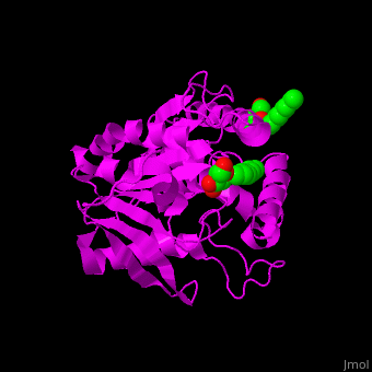
The 3D structures of the 3 antigens show that their active sites are virtually identical indicating that they share the same substrate. However, residues at the surface of the enzymes are different. | The 3D structures of the 3 antigens show that their active sites are virtually identical indicating that they share the same substrate. However, residues at the surface of the enzymes are different. | ||
| - | *<scene name='70/706736/Cv/ | + | *<scene name='70/706736/Cv/5'>Click here to see active site</scene>.<ref>PMID:15192106</ref> |
</StructureSection> | </StructureSection> | ||
Revision as of 10:43, 8 May 2018
| |||||||||||
3D Structures of antigen 85
Updated on 08-May-2018
References
- ↑ Ronning DR, Vissa V, Besra GS, Belisle JT, Sacchettini JC. Mycobacterium tuberculosis antigen 85A and 85C structures confirm binding orientation and conserved substrate specificity. J Biol Chem. 2004 Aug 27;279(35):36771-7. Epub 2004 Jun 10. PMID:15192106 doi:http://dx.doi.org/10.1074/jbc.M400811200
