Glucose transport protein
From Proteopedia
(Difference between revisions)
(New page: ==Your Heading Here (maybe something like 'Structure')== <StructureSection load='1stp' size='340' side='right' caption='Caption for this structure' scene=''> This is a default text for you...) |
|||
| Line 14: | Line 14: | ||
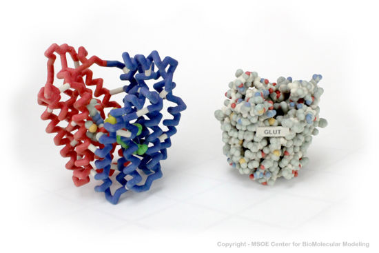
Shown below are 3D printed physical models of the Glucose Transport Protein (GLUT). The backbone model on the left is colored by repeat regions, with the first half red and the second half blue. The spacefill model on the right is colored by atom type, with carbon gray, oxygen red, nitrogen blue and sulfur yellow. | Shown below are 3D printed physical models of the Glucose Transport Protein (GLUT). The backbone model on the left is colored by repeat regions, with the first half red and the second half blue. The spacefill model on the right is colored by atom type, with carbon gray, oxygen red, nitrogen blue and sulfur yellow. | ||
| - | [[Image:glut1_centerForBioMolecularModeling.jpg]] | + | [[Image:glut1_centerForBioMolecularModeling.jpg|550px]] |
====The MSOE Center for BioMolecular Modeling==== | ====The MSOE Center for BioMolecular Modeling==== | ||
Revision as of 20:43, 10 May 2018
Your Heading Here (maybe something like 'Structure')
| |||||||||||
References
- ↑ Hanson, R. M., Prilusky, J., Renjian, Z., Nakane, T. and Sussman, J. L. (2013), JSmol and the Next-Generation Web-Based Representation of 3D Molecular Structure as Applied to Proteopedia. Isr. J. Chem., 53:207-216. doi:http://dx.doi.org/10.1002/ijch.201300024
- ↑ Herraez A. Biomolecules in the computer: Jmol to the rescue. Biochem Mol Biol Educ. 2006 Jul;34(4):255-61. doi: 10.1002/bmb.2006.494034042644. PMID:21638687 doi:10.1002/bmb.2006.494034042644


