We apologize for Proteopedia being slow to respond. For the past two years, a new implementation of Proteopedia has been being built. Soon, it will replace this 18-year old system. All existing content will be moved to the new system at a date that will be announced here.
User:Ricardo Alberto Chiong Zevallos/Sandbox 1
From Proteopedia
(Difference between revisions)
| Line 1: | Line 1: | ||
| - | <StructureSection load=' | + | <StructureSection load='' size='350' side='right' caption='Structure of a Bmi1 protein' scene='78/787701/Bmi1_isolada_da_2h0d/2'> |
| Line 39: | Line 39: | ||
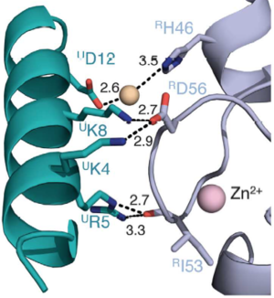
Canonical PRC1 such as BMI1/RING1b have intrinsically very low enzymatic activity compared with non-canonical PRC1, although there are only subtle differences between the structure of canonical and non-canonical complexes. Two charged helix alpha 3 residues present in a modeled BMI1 seems responsible for the low activity of BMI1/RING1b, K73 and D77 form a salt bridge that may limit efficient ubiquitin transfer. In a computational modeling, BMI1 K73 clashes sterically with Ubiquitin, which implies that K73 must move to allow Ub to bind in an activated conformation or that Ub adopts a less optimal position during monoubiquitination. Either of these events could provide an energy barrier, slowing transfer catalyzed by BMI1. The intrinsically low activity of the BMI1/RING1b is offset by a relatively favorable interaction between E3–E2-Ub and nucleosome substrate, resulting in a site-specific monoubiquitination efficient enough. The energy barrier may be responsible for increasing the fidelity of the transfer to the appropriate substrate. Also, canonical and non-canonical differ in targeting sub-units, target genomic loci, and genes expression regulation. | Canonical PRC1 such as BMI1/RING1b have intrinsically very low enzymatic activity compared with non-canonical PRC1, although there are only subtle differences between the structure of canonical and non-canonical complexes. Two charged helix alpha 3 residues present in a modeled BMI1 seems responsible for the low activity of BMI1/RING1b, K73 and D77 form a salt bridge that may limit efficient ubiquitin transfer. In a computational modeling, BMI1 K73 clashes sterically with Ubiquitin, which implies that K73 must move to allow Ub to bind in an activated conformation or that Ub adopts a less optimal position during monoubiquitination. Either of these events could provide an energy barrier, slowing transfer catalyzed by BMI1. The intrinsically low activity of the BMI1/RING1b is offset by a relatively favorable interaction between E3–E2-Ub and nucleosome substrate, resulting in a site-specific monoubiquitination efficient enough. The energy barrier may be responsible for increasing the fidelity of the transfer to the appropriate substrate. Also, canonical and non-canonical differ in targeting sub-units, target genomic loci, and genes expression regulation. | ||
| - | (fig destacando os resíduos K73 and D77 de BMI1 no complexo PRC1) | ||
The central domain of BMI1 forms an <scene name='78/787701/5fr6_bmi1/2'>ubiquitin-like (UBL) domain</scene>, which is involved in protein-protein interactions, including interactions with the transcription factors E4F1, Zfp277 and the PLZF-RARA fusion protein. The best characterized binding partners of the UBL domain are the polyhomeotic proteins (PHC1, PHC2, PHC3). The UBL domain binds a short, 24 amino acid fragment, of PHC2 in a b-hairpin conformation. Also, UBL domain is involved in homo-oligomerization of BMI1. NMR and carbon detected NMR found that residues 30-51 are strongly conserved between PHC2, PHC1 and PHC3 suggesting that BMI1 interacts with the three members of the polyhomeotic family in a very similar manner and with similar affinities. Deletion of the corresponding motif abolished the interaction with BMI1. In the <scene name='78/787701/2na1/2'>PHC2-BMI1 complex</scene>, PHC2 residues 33–47 adopt a <scene name='78/787701/2na1_betahairpin_highlighted/1'>beta-hairpin conformation</scene> in the complex, in greenyellow. The PHC2-BMI1 interaction involves an <scene name='78/787701/2na1_bhairpin_b2_highlighted/1'>antiparallel b-sheet</scene> formed between the beta-hairpin of PHC2 and the beta 2 strand of BMI1 UBL, in magenta. The antiparallel b-sheet is stabilized by the hydrogen bonds between BMI1 Tyr163 and PHC2 Gly46. | The central domain of BMI1 forms an <scene name='78/787701/5fr6_bmi1/2'>ubiquitin-like (UBL) domain</scene>, which is involved in protein-protein interactions, including interactions with the transcription factors E4F1, Zfp277 and the PLZF-RARA fusion protein. The best characterized binding partners of the UBL domain are the polyhomeotic proteins (PHC1, PHC2, PHC3). The UBL domain binds a short, 24 amino acid fragment, of PHC2 in a b-hairpin conformation. Also, UBL domain is involved in homo-oligomerization of BMI1. NMR and carbon detected NMR found that residues 30-51 are strongly conserved between PHC2, PHC1 and PHC3 suggesting that BMI1 interacts with the three members of the polyhomeotic family in a very similar manner and with similar affinities. Deletion of the corresponding motif abolished the interaction with BMI1. In the <scene name='78/787701/2na1/2'>PHC2-BMI1 complex</scene>, PHC2 residues 33–47 adopt a <scene name='78/787701/2na1_betahairpin_highlighted/1'>beta-hairpin conformation</scene> in the complex, in greenyellow. The PHC2-BMI1 interaction involves an <scene name='78/787701/2na1_bhairpin_b2_highlighted/1'>antiparallel b-sheet</scene> formed between the beta-hairpin of PHC2 and the beta 2 strand of BMI1 UBL, in magenta. The antiparallel b-sheet is stabilized by the hydrogen bonds between BMI1 Tyr163 and PHC2 Gly46. | ||
| - | |||
| - | (fig destacando as pontes de hidrogenio entre BMI1 Tyr163 e PHC2 Gly46, da beta-hairpin da PHC2 e folha beta 2 da BMI1 UBL) | ||
MEL18, BMI1 homolog, also binds to PHC2, but with slightly reduced affinity. All BMI1 residues involved in contacts with PHC2 are identical with MEL18 and strongly suggests very similar binding mode for MEL18–PHC2. The similarity is explained by the 60% sequence identity between UBL domain and MEL18. MEL18 is capable of biding to RING1b to form a PRC1 complex too, but the MEL18/RING1b complex is significantly less active than the BMI1/RING1b complex. | MEL18, BMI1 homolog, also binds to PHC2, but with slightly reduced affinity. All BMI1 residues involved in contacts with PHC2 are identical with MEL18 and strongly suggests very similar binding mode for MEL18–PHC2. The similarity is explained by the 60% sequence identity between UBL domain and MEL18. MEL18 is capable of biding to RING1b to form a PRC1 complex too, but the MEL18/RING1b complex is significantly less active than the BMI1/RING1b complex. | ||
| Line 52: | Line 49: | ||
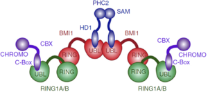
Proposed architecture of the PRC1 complex oligomer. | Proposed architecture of the PRC1 complex oligomer. | ||
| + | |||
| + | |||
| + | |||
| + | == Disease == | ||
| + | |||
| + | The bmi1 gene was first associated with lymphoma, in cooperation with the oncogene myc. BMI1-deficient fibroblasts and lymphocytes show high expression of the tumour suppressors p16 and p19Arf, which are encoded by ink4a locus. Conversely, overexpression of bmi1 allows fibroblast immortalization, downregulates expression of p16 and p19Arf and, leading to neoplastic transformation. The modulation of the ink4a expression probably is due to the ubiquitination of a H2A in the region, compacting the chromatin and downregulating the expression of p16 and p19Arf. The B- and T-cell lymphomas are highly dose-dependent phenotypes regarding bmi1 gene expression. The overexpression can lead to B- and T-cell lymphomas and partial reduction of Bmi1 leads to significant reduction in lymphoma formation and brain tumour formation. | ||
| + | |||
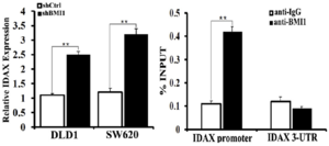
| + | BMI1 is also associated with colon cancer, in which BMI1 activates WNT signaling mainly through the downregulation of IDAX expression. The BMI1 protein binds to the promoter of IDAX, a Wnt antagonist, and decreases it’s transcription. The WNT signaling pathway is very important in cell fate determination and tissue development and stem cell maintenance. Excessive activation of this pathway leads to tumorigenesis in several types of human cancers. According to The Cancer Genome Atlas Network, the WNT pathway is hyper-activated in over 90% of colon cancer. | ||
| + | |||
| + | [[Image:BMI1 regulates IDAX transcription.png|300px]] | ||
| + | |||
| + | IDAX as a target from BMI1. In the left is shown that the BMI1 knockdown by short hairpin RNAs leads to the increased IDAX transcription in colon cancer cell lines. In the right is shown the binding of the BMI1 protein to the promoter of IDAX, analyzed by ChIP-PCR. | ||
| + | |||
| + | BMI1 can bind to the promoter of HoxC13 gene to regulate it and also can regulate other Hox genes, which define axial patterning and segment identity. BMI1 also inhibits the CDKN1A gene, an important inhibitor of the cellular cycle, promoting the cellular proliferation. The inhibition of CDKN1A gene is promoted when a lncRNA, FALEC (focally amplified long non-coding RNA on chromosome 1 gene), stabilizes BMI1. | ||
| + | |||
| + | |||
| + | |||
== Conservation == | == Conservation == | ||
| Line 64: | Line 78: | ||
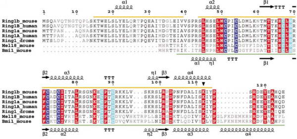
Sequence alignment of Ring-domain proteins in PRC1 with secondary structure indicated. Zn binding site I is highlighted in blue and Zn binding site II is highlighted in cyan. | Sequence alignment of Ring-domain proteins in PRC1 with secondary structure indicated. Zn binding site I is highlighted in blue and Zn binding site II is highlighted in cyan. | ||
| + | [[Image:Consurf_key_small.gif|200px|right]] | ||
| + | Check<jmol> | ||
| + | <jmolCheckbox> | ||
| + | <scriptWhenChecked>select protein; define ~consurf_to_do selected; consurf_initial_scene = true; script "/wiki/ConSurf/h0/2h0d_consurf.spt"</scriptWhenChecked> | ||
| + | <scriptWhenUnchecked>script /wiki/extensions/Proteopedia/spt/initialview01.spt</scriptWhenUnchecked> | ||
| + | <text>to colour the structure by Evolutionary Conservation</text> | ||
| + | </jmolCheckbox> | ||
| + | </jmol>, as determined by [http://consurfdb.tau.ac.il/ ConSurfDB]. You may read the [[Conservation%2C_Evolutionary|explanation]] of the method and the full data available from [http://bental.tau.ac.il/new_ConSurfDB/main_output.php?pdb_ID=2h0d ConSurf]. | ||
| + | <div style="clear:both"></div> | ||
| + | <div style="background-color:#fffaf0;"> | ||
| - | == Disease == | ||
| - | |||
| - | The bmi1 gene was first associated with lymphoma, in cooperation with the oncogene myc. BMI1-deficient fibroblasts and lymphocytes show high expression of the tumour suppressors p16 and p19Arf, which are encoded by ink4a locus. Conversely, overexpression of bmi1 allows fibroblast immortalization, downregulates expression of p16 and p19Arf and, leading to neoplastic transformation. The modulation of the ink4a expression probably is due to the ubiquitination of a H2A in the region, compacting the chromatin and downregulating the expression of p16 and p19Arf. The B- and T-cell lymphomas are highly dose-dependent phenotypes regarding bmi1 gene expression. The overexpression can lead to B- and T-cell lymphomas and partial reduction of Bmi1 leads to significant reduction in lymphoma formation and brain tumour formation. | ||
| - | |||
| - | BMI1 is also associated with colon cancer, in which BMI1 activates WNT signaling mainly through the downregulation of IDAX expression. The BMI1 protein binds to the promoter of IDAX, a Wnt antagonist, and decreases it’s transcription. The WNT signaling pathway is very important in cell fate determination and tissue development and stem cell maintenance. Excessive activation of this pathway leads to tumorigenesis in several types of human cancers. According to The Cancer Genome Atlas Network, the WNT pathway is hyper-activated in over 90% of colon cancer. | ||
| - | |||
| - | [[Image:BMI1 regulates IDAX transcription.png]] | ||
| - | |||
| - | IDAX as a target from BMI1. In the left is shown that the BMI1 knockdown by short hairpin RNAs leads to the increased IDAX transcription in colon cancer cell lines. In the right is shown the binding of the BMI1 protein to the promoter of IDAX, analyzed by ChIP-PCR. | ||
| - | |||
| - | BMI1 can bind to the promoter of HoxC13 gene to regulate it and also can regulate other Hox genes, which define axial patterning and segment identity. BMI1 also inhibits the CDKN1A gene, an important inhibitor of the cellular cycle, promoting the cellular proliferation. The inhibition of CDKN1A gene is promoted when a lncRNA, FALEC (focally amplified long non-coding RNA on chromosome 1 gene), stabilizes BMI1. | ||
</StructureSection> | </StructureSection> | ||
Revision as of 21:14, 17 June 2018
| |||||||||||
References
- ↑ Jacobs JJ, Kieboom K, Marino S, DePinho RA, van Lohuizen M. The oncogene and Polycomb-group gene bmi-1 regulates cell proliferation and senescence through the ink4a locus. Nature. 1999 Jan 14;397(6715):164-8. doi: 10.1038/16476. PMID:9923679 doi:http://dx.doi.org/10.1038/16476
- ↑ Wang H, Wang L, Erdjument-Bromage H, Vidal M, Tempst P, Jones RS, Zhang Y. Role of histone H2A ubiquitination in Polycomb silencing. Nature. 2004 Oct 14;431(7010):873-8. Epub 2004 Sep 22. PMID:15386022 doi:10.1038/nature02985
- ↑ Gray F, Cho HJ, Shukla S, He S, Harris A, Boytsov B, Jaremko L, Jaremko M, Demeler B, Lawlor ER, Grembecka J, Cierpicki T. BMI1 regulates PRC1 architecture and activity through homo- and hetero-oligomerization. Nat Commun. 2016 Nov 9;7:13343. doi: 10.1038/ncomms13343. PMID:27827373 doi:http://dx.doi.org/10.1038/ncomms13343
- ↑ Bentley ML, Corn JE, Dong KC, Phung Q, Cheung TK, Cochran AG. Recognition of UbcH5c and the nucleosome by the Bmi1/Ring1b ubiquitin ligase complex. EMBO J. 2011 Jul 19. doi: 10.1038/emboj.2011.243. PMID:21772249 doi:10.1038/emboj.2011.243
- ↑ Taherbhoy AM, Huang OW, Cochran AG. BMI1-RING1B is an autoinhibited RING E3 ubiquitin ligase. Nat Commun. 2015 Jul 7;6:7621. doi: 10.1038/ncomms8621. PMID:26151332 doi:http://dx.doi.org/10.1038/ncomms8621
https://drive.google.com/drive/folders/1l195aNuY6joOd74GKKxa-XWTRMBv_uWF?usp=sharing


