User:Kayque Alves Telles Silva/Sandbox 1
From Proteopedia
(Difference between revisions)
| Line 20: | Line 20: | ||
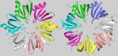
The proximal face of Hfq interacts with U-rich 5′-AUUUUUG-3′ RNA sequences, forming a small <scene name='78/789833/Hfq-rna/2'>ring of RNA</scene> around its pore. Residues such as '''Tyr42''', '''Lys41''' and '''Gln8''' guarantee the bond with uracils and each subunit contacts one nucleotide, except the RNA guanine that remains exposed out of the ring. However, in the predominantly <scene name='78/789833/Rna_distal_face/2'>hydrophobic distal face</scene>, each subunit bonds with three nucleotides, forming a wider ring of RNA. In this case, A(A/G)N - N being any base - sequences are preferred. | The proximal face of Hfq interacts with U-rich 5′-AUUUUUG-3′ RNA sequences, forming a small <scene name='78/789833/Hfq-rna/2'>ring of RNA</scene> around its pore. Residues such as '''Tyr42''', '''Lys41''' and '''Gln8''' guarantee the bond with uracils and each subunit contacts one nucleotide, except the RNA guanine that remains exposed out of the ring. However, in the predominantly <scene name='78/789833/Rna_distal_face/2'>hydrophobic distal face</scene>, each subunit bonds with three nucleotides, forming a wider ring of RNA. In this case, A(A/G)N - N being any base - sequences are preferred. | ||
| - | Interestingly, the RNA binding promotes a conformational change in Hfq, highlighted by the pore diameter enlargement. The native protein shows a pore of roughly <scene name='78/789833/Hfq_pore_small/ | + | Interestingly, the RNA binding promotes a conformational change in Hfq, highlighted by the pore diameter enlargement. The native protein shows a pore of roughly <scene name='78/789833/Hfq_pore_small/2'>12Å</scene>, but in case of active chaperone activity, the RNA bond promotes a pore diameter deformation to <scene name='78/789833/Hfq_pore_large/3'>15Å</scene>. |
== Structural highlights == | == Structural highlights == | ||
Revision as of 04:58, 18 June 2018
Hfq
| |||||||||||
References
- ↑ Hanson, R. M., Prilusky, J., Renjian, Z., Nakane, T. and Sussman, J. L. (2013), JSmol and the Next-Generation Web-Based Representation of 3D Molecular Structure as Applied to Proteopedia. Isr. J. Chem., 53:207-216. doi:http://dx.doi.org/10.1002/ijch.201300024
- ↑ Herraez A. Biomolecules in the computer: Jmol to the rescue. Biochem Mol Biol Educ. 2006 Jul;34(4):255-61. doi: 10.1002/bmb.2006.494034042644. PMID:21638687 doi:10.1002/bmb.2006.494034042644

