User:Kayque Alves Telles Silva/Sandbox 1
From Proteopedia
(Difference between revisions)
| Line 1: | Line 1: | ||
==Hfq== | ==Hfq== | ||
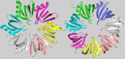
<StructureSection load='1kq1' size='400' side='right' caption="Host Factor for the Replication of the Qβ Phage RNA (Hfq)"> | <StructureSection load='1kq1' size='400' side='right' caption="Host Factor for the Replication of the Qβ Phage RNA (Hfq)"> | ||
| - | Hfq is a bacterial cytoplasmatic protein, first named for being a Host Factor for the Replication of the Qβ Phage RNA<ref>DOI 10.1093/emboj/cdf322</ref>. Hfq acts pleiotropically and impacts both the degradation and the translation efficiency of mRNAs<ref>DOI 10.1046/j.1365-2443.2000.00350.x</ref>, through the modification of sRNA | + | Hfq is a bacterial cytoplasmatic protein, first named for being a Host Factor for the Replication of the Qβ Phage RNA<ref>DOI 10.1093/emboj/cdf322</ref>. Hfq acts pleiotropically and impacts both the degradation and the translation efficiency of mRNAs<ref>DOI 10.1046/j.1365-2443.2000.00350.x</ref>, through the modification of sRNA<ref>DOI 10.1101/gad.901001</ref>. Besides, Hfq has a strongly conserved structure[9] and belongs to the LSm protein family, present in each of the three domains of life. Despite Hfq’s underscored importance, there is a wide spectrum of cellular functions, as growth rate and cell length, that are directly dependent of its chaperone function and have been discovered in the last century<ref>DOI 10.1016/j.mib.2016.02.003</ref><ref>DOI 10.1002/wrna.1475</ref><ref>DOI 10.15252/embj.201797631</ref>, but many others remain unknown. |
== Function == | == Function == | ||
| - | <scene name='78/789833/Hfq/2'>Hfq</scene> is a doughnut shaped homohexamer of roughly 8,9kDa, ~65Å diameter and ~23Å width, as described in S.aureus[1]. The action mechanisms of Hfq as a chaperone are based in its interaction with sRNAs. The best literature described functions among them are: the sequestering of ribosome binding sites (RBS); the inhibition of RBS-blocking mRNA regions, allowing translation; the stabilization of sRNAs which would otherwise be degraded; and the degradation of mRNAs. Hfq might also contribute to the degradation of certain mRNAs directly, but rarely[ | + | <scene name='78/789833/Hfq/2'>Hfq</scene> is a doughnut shaped homohexamer of roughly 8,9kDa, ~65Å diameter and ~23Å width, as described in S.aureus[1]. The action mechanisms of Hfq as a chaperone are based in its interaction with sRNAs. The best literature described functions among them are: the sequestering of ribosome binding sites (RBS); the inhibition of RBS-blocking mRNA regions, allowing translation; the stabilization of sRNAs which would otherwise be degraded; and the degradation of mRNAs. Hfq might also contribute to the degradation of certain mRNAs directly, but rarely[7]. |
== Structure == | == Structure == | ||
| - | The <scene name='78/789833/Subunit/1'>basic subunit</scene> which forms the homo-hexamer of Hfq has 77 residues[1] comprising one N-terminal α helix (α1), five beta sheets (β1-β5) and one variable region. An unstructured carboxy-terminal region, which may have >100 residues in certain species is also present[ | + | The <scene name='78/789833/Subunit/1'>basic subunit</scene> which forms the homo-hexamer of Hfq has 77 residues[1] comprising one N-terminal α helix (α1), five beta sheets (β1-β5) and one variable region. An unstructured carboxy-terminal region, which may have >100 residues in certain species is also present[8]. The fold of the beta sheets is characterized by two important <scene name='78/789833/Motifs/5'>motifs</scene>, Sm1 and Sm2, used to clusterize the LSm protein family components considering their highlighted conservation throughout Life’s domains. |
The most conserved amino acids of Hfq are the aspartic acid 40 and the glycine 34, determining hydrophobic residues present in the Sm1 motif which maintain the highly distorted Sm1 fold. Tyr56 and Tyr63, highly conserved in the Sm2 motif, are fundamental for the interaction between subunits. The glutamine 8 and tyrosine 42 are highly conserved in Hfq proteins due to their role in uracil binding[1]. The highly variable C-terminus of Hfq proteins has no defined structure and its function is not clearly understood yet[9]. | The most conserved amino acids of Hfq are the aspartic acid 40 and the glycine 34, determining hydrophobic residues present in the Sm1 motif which maintain the highly distorted Sm1 fold. Tyr56 and Tyr63, highly conserved in the Sm2 motif, are fundamental for the interaction between subunits. The glutamine 8 and tyrosine 42 are highly conserved in Hfq proteins due to their role in uracil binding[1]. The highly variable C-terminus of Hfq proteins has no defined structure and its function is not clearly understood yet[9]. | ||
Revision as of 05:13, 18 June 2018
Hfq
| |||||||||||
References
- ↑ Schumacher MA, Pearson RF, Moller T, Valentin-Hansen P, Brennan RG. Structures of the pleiotropic translational regulator Hfq and an Hfq-RNA complex: a bacterial Sm-like protein. EMBO J. 2002 Jul 1;21(13):3546-56. PMID:12093755 doi:10.1093/emboj/cdf322
- ↑ doi: https://dx.doi.org/10.1046/j.1365-2443.2000.00350.x
- ↑ Wassarman KM, Repoila F, Rosenow C, Storz G, Gottesman S. Identification of novel small RNAs using comparative genomics and microarrays. Genes Dev. 2001 Jul 1;15(13):1637-51. doi: 10.1101/gad.901001. PMID:11445539 doi:http://dx.doi.org/10.1101/gad.901001
- ↑ Updegrove TB, Zhang A, Storz G. Hfq: the flexible RNA matchmaker. Curr Opin Microbiol. 2016 Apr;30:133-138. doi: 10.1016/j.mib.2016.02.003. Epub, 2016 Feb 22. PMID:26907610 doi:http://dx.doi.org/10.1016/j.mib.2016.02.003
- ↑ Santiago-Frangos A, Woodson SA. Hfq chaperone brings speed dating to bacterial sRNA. Wiley Interdiscip Rev RNA. 2018 Jul;9(4):e1475. doi: 10.1002/wrna.1475. Epub 2018, Apr 6. PMID:29633565 doi:http://dx.doi.org/10.1002/wrna.1475
- ↑ Andrade JM, Dos Santos RF, Chelysheva I, Ignatova Z, Arraiano CM. The RNA-binding protein Hfq is important for ribosome biogenesis and affects translation fidelity. EMBO J. 2018 Jun 1;37(11). pii: embj.201797631. doi: 10.15252/embj.201797631., Epub 2018 Apr 18. PMID:29669858 doi:http://dx.doi.org/10.15252/embj.201797631
- ↑ Hanson, R. M., Prilusky, J., Renjian, Z., Nakane, T. and Sussman, J. L. (2013), JSmol and the Next-Generation Web-Based Representation of 3D Molecular Structure as Applied to Proteopedia. Isr. J. Chem., 53:207-216. doi:http://dx.doi.org/10.1002/ijch.201300024
- ↑ Herraez A. Biomolecules in the computer: Jmol to the rescue. Biochem Mol Biol Educ. 2006 Jul;34(4):255-61. doi: 10.1002/bmb.2006.494034042644. PMID:21638687 doi:10.1002/bmb.2006.494034042644

