We apologize for Proteopedia being slow to respond. For the past two years, a new implementation of Proteopedia has been being built. Soon, it will replace this 18-year old system. All existing content will be moved to the new system at a date that will be announced here.
Polygalacturonase
From Proteopedia
(Difference between revisions)
| Line 1: | Line 1: | ||
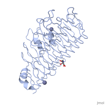
| - | < | + | <StructureSection load='1czf' size='350' side='right' caption='Glycosylated structure of endo-polygalacturonase II complex with Zn+2 ion (grey) (PDB code [[1czf]])' scene=''> |
'''Polygalacturonases''' (PGs) catalyze the enzymatic depolymerization of pectates – polysaccharides that comprise the plant cell wall. Polymer disassembly of substrates by ''exo-'' and ''endo-'' PGs is carried out via a hydrolytic mechanism. Degradation of pectates in plant cell walls contributes to ripening of fruits, such as tomatoes and melons<ref>PMID:9625687</ref>. Microbial PGs have been identified to be a part of defense mechanisms because of their role in pathogen attack<ref name="crystal">PMID:9733763</ref>. | '''Polygalacturonases''' (PGs) catalyze the enzymatic depolymerization of pectates – polysaccharides that comprise the plant cell wall. Polymer disassembly of substrates by ''exo-'' and ''endo-'' PGs is carried out via a hydrolytic mechanism. Degradation of pectates in plant cell walls contributes to ripening of fruits, such as tomatoes and melons<ref>PMID:9625687</ref>. Microbial PGs have been identified to be a part of defense mechanisms because of their role in pathogen attack<ref name="crystal">PMID:9733763</ref>. | ||
| Line 21: | Line 21: | ||
Not all PGs have ten coils in the parallel beta helix. They usually have approximately 10 coils. [[Image:PBH2.jpg|200px|left|thumb| Nomenclature for structural elements of the parallel beta helix<ref name="yoder" />. PB1 is Parallel Beta Sheet 1, T1 is Turn 1, between PB1 and PB2., [[PBH2]]]] With the parallel beta helix fold, the three major beta sheets are call PB1, PB2, and PB3. The turns between strands are Turn 1 (T1) between PB1 and PB2, T2 is the turn between PB2 and PB3, and T3 is the turn between PB3 and PB1 of the next coil. For your reference, this is illustrated in the figure below. | Not all PGs have ten coils in the parallel beta helix. They usually have approximately 10 coils. [[Image:PBH2.jpg|200px|left|thumb| Nomenclature for structural elements of the parallel beta helix<ref name="yoder" />. PB1 is Parallel Beta Sheet 1, T1 is Turn 1, between PB1 and PB2., [[PBH2]]]] With the parallel beta helix fold, the three major beta sheets are call PB1, PB2, and PB3. The turns between strands are Turn 1 (T1) between PB1 and PB2, T2 is the turn between PB2 and PB3, and T3 is the turn between PB3 and PB1 of the next coil. For your reference, this is illustrated in the figure below. | ||
| + | |||
</StructureSection> | </StructureSection> | ||
Revision as of 09:42, 23 August 2018
| |||||||||||

3D Structures of polygalacturonase
Updated on 23-August-2018
References
- ↑ Hadfield KA, Bennett AB. Polygalacturonases: many genes in search of a function. Plant Physiol. 1998 Jun;117(2):337-43. PMID:9625687
- ↑ 2.0 2.1 2.2 Pickersgill R, Smith D, Worboys K, Jenkins J. Crystal structure of polygalacturonase from Erwinia carotovora ssp. carotovora. J Biol Chem. 1998 Sep 18;273(38):24660-4. PMID:9733763
- ↑ 3.0 3.1 Yoder MD, Lietzke SE, Jurnak F. Unusual structural features in the parallel beta-helix in pectate lyases. Structure. 1993 Dec 15;1(4):241-51. PMID:8081738
Proteopedia Page Contributors and Editors (what is this?)
Joel L. Sussman, Krishna Amin, Michal Harel, Marilyn Yoder, OCA, Jaime Prilusky
