This old version of Proteopedia is provided for student assignments while the new version is undergoing repairs. Content and edits done in this old version of Proteopedia after March 1, 2026 will eventually be lost when it is retired in about June of 2026.
Apply for new accounts at the new Proteopedia. Your logins will work in both the old and new versions.
Xanthine dehydrogenase
From Proteopedia
(Difference between revisions)
| Line 4: | Line 4: | ||
Several co-crystal structures of '''xanthine oxidase''' and '''xanthine dehydrogenase''' (which do not differ in conformation of active site) were studied to understand key interactions for enzyme inhibition. Docking of novel hits and inactive compounds was performed (to <scene name='Journal:BMC:3/Cv/3'>protein</scene> in PDB with ID [[1vdv]]) to understand the ligand-protein interactions and hence, for structure-based design of more potent molecules.<ref >doi 10.1016/j.bmc.2012.03.019</ref> The paper reports the directions for modification of the hit compound derived from these considerations, which are reported below. The mechanism of action of the novel hits are like <scene name='Journal:BMC:3/Cv/6'>piraxostat</scene> and <scene name='Journal:BMC:3/Cv/5'>febuxostat</scene> (“pure inhibitors”) and not like <scene name='Journal:BMC:3/Cv/8'>allopurinol</scene> & '''FYX-051''' (“substrate inhibitors”). | Several co-crystal structures of '''xanthine oxidase''' and '''xanthine dehydrogenase''' (which do not differ in conformation of active site) were studied to understand key interactions for enzyme inhibition. Docking of novel hits and inactive compounds was performed (to <scene name='Journal:BMC:3/Cv/3'>protein</scene> in PDB with ID [[1vdv]]) to understand the ligand-protein interactions and hence, for structure-based design of more potent molecules.<ref >doi 10.1016/j.bmc.2012.03.019</ref> The paper reports the directions for modification of the hit compound derived from these considerations, which are reported below. The mechanism of action of the novel hits are like <scene name='Journal:BMC:3/Cv/6'>piraxostat</scene> and <scene name='Journal:BMC:3/Cv/5'>febuxostat</scene> (“pure inhibitors”) and not like <scene name='Journal:BMC:3/Cv/8'>allopurinol</scene> & '''FYX-051''' (“substrate inhibitors”). | ||
| - | Xanthine oxidoreductase (XOR), is an oxidoreductive enzyme that is synthesized as '''xanthine dehydrogenase (XDH)''' and can be converted reversibly or irreversibly to xanthine oxidase (XO) form. It catalyzes the <scene name='Journal:BMC:3/Cv/13'>transformation of physiological substrates</scene> such as <scene name='Journal:BMC:3/Cv/16'>hypoxanthine to xanthine</scene> and <scene name='Journal:BMC:3/Cv/17'>xanthine to uric acid</scene> which is excreted by kidneys.<ref name="Pauff">PMID:19109252</ref> The reaction occurs at the <scene name='Journal:BMC:3/Cv2/5'>cofactor molybdopterin (Mo-Pt)</scene> center from where the <scene name='Journal:BMC:3/Cv2/6'>electrons are transferred via two Fe2S2 clusters to FAD, which then passes them on to the second substrate NAD+</scene> in case of XDH (PDB code [[2w3s]])<ref name="Xan">PMID: 19109249</ref> or to molecular oxygen in XO leading to the formation of superoxide anion and H<sub>2</sub>O<sub>2</sub>. Excessive production and/or inadequate excretion of uric acid results in hyperuricemia and is associated with conditions like gout, cardiovascular mortality and metabolic syndrome including hyperinsulinemia and hypertriglyceridemia. Alleviating hyperuricemia, therefore, has therapeutic significance, and XO is a key target towards this end. | + | '''Xanthine oxidoreductase''' (XOR), is an oxidoreductive enzyme that is synthesized as '''xanthine dehydrogenase (XDH)''' and can be converted reversibly or irreversibly to xanthine oxidase (XO) form. It catalyzes the <scene name='Journal:BMC:3/Cv/13'>transformation of physiological substrates</scene> such as <scene name='Journal:BMC:3/Cv/16'>hypoxanthine to xanthine</scene> and <scene name='Journal:BMC:3/Cv/17'>xanthine to uric acid</scene> which is excreted by kidneys.<ref name="Pauff">PMID:19109252</ref> The reaction occurs at the <scene name='Journal:BMC:3/Cv2/5'>cofactor molybdopterin (Mo-Pt)</scene> center from where the <scene name='Journal:BMC:3/Cv2/6'>electrons are transferred via two Fe2S2 clusters to FAD, which then passes them on to the second substrate NAD+</scene> in case of XDH (PDB code [[2w3s]])<ref name="Xan">PMID: 19109249</ref> or to molecular oxygen in XO leading to the formation of superoxide anion and H<sub>2</sub>O<sub>2</sub>. Excessive production and/or inadequate excretion of uric acid results in hyperuricemia and is associated with conditions like gout, cardiovascular mortality and metabolic syndrome including hyperinsulinemia and hypertriglyceridemia. Alleviating hyperuricemia, therefore, has therapeutic significance, and XO is a key target towards this end. |
Piraxostat (PDB code [[1vdv]]) <ref name="Fukunari">PMID: 15190124</ref> and febuxostat (PDB code [[1n5x]])<ref name="Okamoto"/>, show several interactions with the active site residues of the protein. The carboxyl group of piraxostat is involved in <scene name='Journal:BMC:3/Cv1/3'>electrostatic interactions with guanidinium group of Arg880</scene> and <scene name='Journal:BMC:3/Cv1/4'>H-bonds to Thr1010</scene> as well. The ring nitrogen is involved in <scene name='Journal:BMC:3/Cv1/5'>H-bond interaction with Glu802</scene>. The cyano group of the ligand forms another <scene name='Journal:BMC:3/Cv1/7'>crucial H-bond with Asn768</scene>. Besides these polar interactions, a number of hydrophobic interactions are observed as well. The heteroaromatic ring is <scene name='Journal:BMC:3/Cv1/8'>pi-stacked between Phe914 and Phe1009</scene>. The phenyl ring has hydrophobic interactions with <scene name='Journal:BMC:3/Cv1/9'>Leu873, Val1011 and Leu1014</scene>. The alkoxy side chain extends towards the solvent accessible region and is engaged in hydrophobic interactions with various residues at the entrance of the pocket such as <scene name='Journal:BMC:3/Cv1/10'>Leu648, Phe649 and Phe1013</scene>. <span style="color:lime;background-color:black;font-weight:bold;">Piraxostat is in green</span>, <span style="color:deepskyblue;background-color:black;font-weight:bold;">Mo-Pt is in deep-sky-blue</span>, residues are colored according to the type of interaction with ligand – <span style="color:salmon;background-color:black;font-weight:bold;">salmon for pi-stack</span>, <font color='magenta'><b>magenta for other hydrophobic</b></font> and <span style="color:cyan;background-color:black;font-weight:bold;">cyan for polar interactions</span>. | Piraxostat (PDB code [[1vdv]]) <ref name="Fukunari">PMID: 15190124</ref> and febuxostat (PDB code [[1n5x]])<ref name="Okamoto"/>, show several interactions with the active site residues of the protein. The carboxyl group of piraxostat is involved in <scene name='Journal:BMC:3/Cv1/3'>electrostatic interactions with guanidinium group of Arg880</scene> and <scene name='Journal:BMC:3/Cv1/4'>H-bonds to Thr1010</scene> as well. The ring nitrogen is involved in <scene name='Journal:BMC:3/Cv1/5'>H-bond interaction with Glu802</scene>. The cyano group of the ligand forms another <scene name='Journal:BMC:3/Cv1/7'>crucial H-bond with Asn768</scene>. Besides these polar interactions, a number of hydrophobic interactions are observed as well. The heteroaromatic ring is <scene name='Journal:BMC:3/Cv1/8'>pi-stacked between Phe914 and Phe1009</scene>. The phenyl ring has hydrophobic interactions with <scene name='Journal:BMC:3/Cv1/9'>Leu873, Val1011 and Leu1014</scene>. The alkoxy side chain extends towards the solvent accessible region and is engaged in hydrophobic interactions with various residues at the entrance of the pocket such as <scene name='Journal:BMC:3/Cv1/10'>Leu648, Phe649 and Phe1013</scene>. <span style="color:lime;background-color:black;font-weight:bold;">Piraxostat is in green</span>, <span style="color:deepskyblue;background-color:black;font-weight:bold;">Mo-Pt is in deep-sky-blue</span>, residues are colored according to the type of interaction with ligand – <span style="color:salmon;background-color:black;font-weight:bold;">salmon for pi-stack</span>, <font color='magenta'><b>magenta for other hydrophobic</b></font> and <span style="color:cyan;background-color:black;font-weight:bold;">cyan for polar interactions</span>. | ||
Revision as of 17:06, 9 October 2018
| |||||||||||
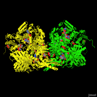
3D structures of xanthine dehydrogenase
Updated on 09-October-2018
+ salicilate + NADH
References
- ↑ B-Rao C, Kulkarni-Almeida A, Katkar KV, Khanna S, Ghosh U, Keche A, Shah P, Srivastava A, Korde V, Nemmani KV, Deshmukh NJ, Dixit A, Brahma MK, Bahirat U, Doshi L, Sharma R, Sivaramakrishnan H. Identification of novel isocytosine derivatives as xanthine oxidase inhibitors from a set of virtual screening hits. Bioorg Med Chem. 2012 May 1;20(9):2930-9. Epub 2012 Mar 14. PMID:22483591 doi:10.1016/j.bmc.2012.03.019
- ↑ Pauff JM, Cao H, Hille R. Substrate Orientation and Catalysis at the Molybdenum Site in Xanthine Oxidase: CRYSTAL STRUCTURES IN COMPLEX WITH XANTHINE AND LUMAZINE. J Biol Chem. 2009 Mar 27;284(13):8760-7. Epub 2008 Dec 24. PMID:19109252 doi:10.1074/jbc.M804517200
- ↑ Dietzel U, Kuper J, Doebbler JA, Schulte A, Truglio JJ, Leimkuhler S, Kisker C. Mechanism of Substrate and Inhibitor Binding of Rhodobacter capsulatus Xanthine Dehydrogenase. J Biol Chem. 2009 Mar 27;284(13):8768-76. Epub 2008 Dec 24. PMID:19109249 doi:http://dx.doi.org/10.1074/jbc.M808114200
- ↑ Fukunari A, Okamoto K, Nishino T, Eger BT, Pai EF, Kamezawa M, Yamada I, Kato N. Y-700 [1-[3-Cyano-4-(2,2-dimethylpropoxy)phenyl]-1H-pyrazole-4-carboxylic acid]: a potent xanthine oxidoreductase inhibitor with hepatic excretion. J Pharmacol Exp Ther. 2004 Nov;311(2):519-28. Epub 2004 Jun 9. PMID:15190124 doi:10.1124/jpet.104.070433
- ↑ 5.0 5.1 Okamoto K, Eger BT, Nishino T, Kondo S, Pai EF, Nishino T. An extremely potent inhibitor of xanthine oxidoreductase. Crystal structure of the enzyme-inhibitor complex and mechanism of inhibition. J Biol Chem. 2003 Jan 17;278(3):1848-55. Epub 2002 Nov 5. PMID:12421831 doi:10.1074/jbc.M208307200
- ↑ Truglio JJ, Theis K, Leimkuhler S, Rappa R, Rajagopalan KV, Kisker C. Crystal structures of the active and alloxanthine-inhibited forms of xanthine dehydrogenase from Rhodobacter capsulatus. Structure. 2002 Jan;10(1):115-25. PMID:11796116
- ↑ 7.0 7.1 Okamoto K, Matsumoto K, Hille R, Eger BT, Pai EF, Nishino T. The crystal structure of xanthine oxidoreductase during catalysis: implications for reaction mechanism and enzyme inhibition. Proc Natl Acad Sci U S A. 2004 May 25;101(21):7931-6. Epub 2004 May 17. PMID:15148401 doi:10.1073/pnas.0400973101
- ↑ Correia HD, Marangon J, Brondino CD, Moura JJ, Romao MJ, Gonzalez PJ, Santos-Silva T. Aromatic aldehydes at the active site of aldehyde oxidoreductase from Desulfovibrio gigas: reactivity and molecular details of the enzyme-substrate and enzyme-product interaction. J Biol Inorg Chem. 2014 Sep 27. PMID:25261288 doi:http://dx.doi.org/10.1007/s00775-014-1196-4
