Main Page

From Proteopedia

(Difference between revisions)
Jump to: navigation, search
Line 35: Line 35:
<td style="padding: 10px;>
<p>[[:Category:Featured in Selected Pages|Other Selected Pages]]</p>
-
<p>[[Help:Contents#For_authors:_contributing_content|How to author pages and contribute to Proteopedia]]</p>
+
<p>[[Help:Contents#For_authors:_contributing_content|How to add content to Proteopedia]]</p>
<p>[[Proteopedia:Video_Guide|Video Guides]]</p>
<p>[[Who knows]] ...</p>

Revision as of 07:08, 21 October 2018

ISSN 2310-6301

Because life has more than 2D, Proteopedia helps to understand relationships between structure and function. Proteopedia is a free, collaborative 3D-encyclopedia of proteins & other molecules.


Selected Pages Art on Science Journals Education
About this image
Avian Influenza Neuraminidase

Eric Martz
The first new influenza virus to emerge as an imminent pandemic threat in the 21st century is H1N1 swine flu. The drug oseltamivir (Tamiflu®) inhibits flu neuraminidase, a component necessary for virus spread, in susceptible flu strains. The development of oseltamivir was guided, in part, by crystallographically determined structures of flu neuraminidase, which is a homotetramer, shown with oseltamivir bound. Oseltamivir was designed to fit N2/N9 (neuraminidases from other strains of flu). Serendipitously, it also fits N1 by induced fit.

>>> Visit this page >>>

About this image
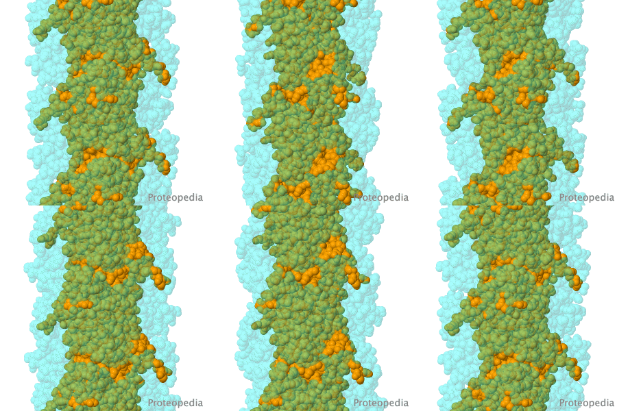
Molecular Sculpture

by Eric Martz
A historical review on sculptures and physical models of macromolecules.

>>> Visit this page >>>

About this image
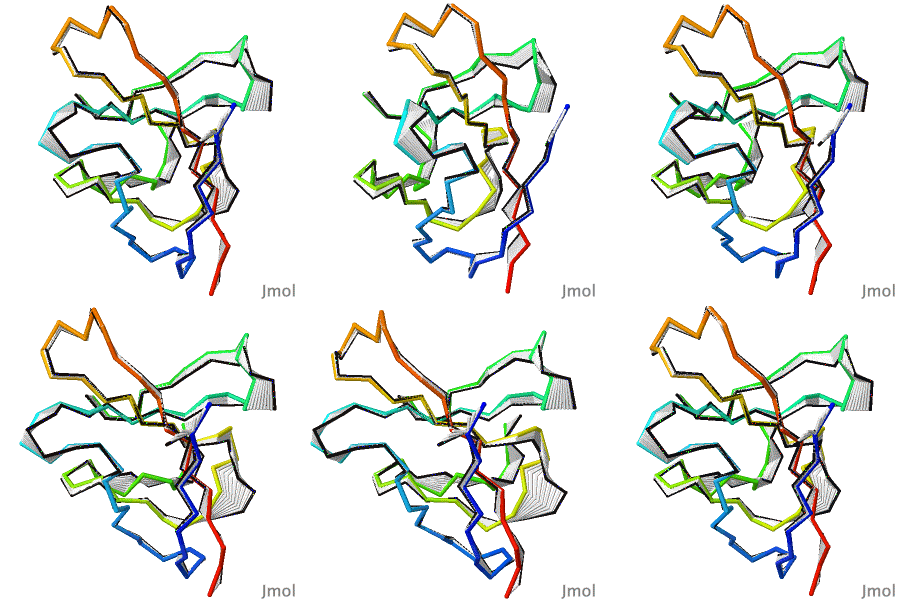
Structural flexibility of the periplasmic protein, FlgA, regulates flagellar P-ring assembly in Salmonella enterica.

H Matsunami, YH Yoon, VA Meshcheryakov, K Namba, FA Samatey. Scientific Reports 2016 doi: 10.1038/srep27399
A periplasmic flagellar chaperone protein, FlgA, is required for P-ring assembly in bacterial flagella of taxa such as Salmonella enterica or Escherichia coli. Here we present the open and closed crystal structures of FlgA from Salmonella enterica serovar Typhimurium, grown under different crystallization conditions. An intramolecular disulfide cross-linked form of FlgA caused a dominant negative effect on motility of the wild-type strain.

>>> Visit this I3DC complement >>>

About this image
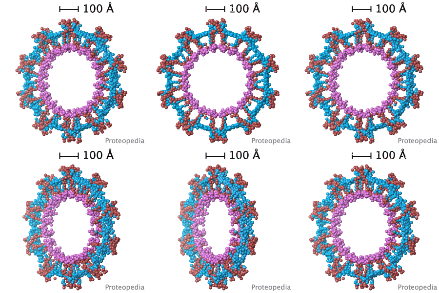
Virus Capsid Geometry

The Capsid of a virus is its outer shell or "skin". Viruses have evolved intricate and elegant ways to assemble capsid protein chains into complete, usually spherical capsids, often with icosahedral symmetry. Pictured is an extremely simplified model of a capsid, where a single enlarged atom represents each of the 360 protein chains in the capsid of the Simian Virus 40 (SV40), a member of a group of cancer-causing viruses that has been extensively researched for decades.

>>> See more animations and explanation >>>

Other Selected Pages

How to add content to Proteopedia

Video Guides

Who knows ...

Featured in Art

All Art on Science

Featured in I3DC

How to get an Interactive 3D Complement for your paper

List of Interactive Complements

About Interactive 3D Complements

Featured in Education

Teaching Strategies Using Proteopedia

Examples of Pages for Teaching

How to author pages and contribute to Proteopedia

About Image:Contact-email.png Table of Contents Structure Index Help

Proteopedia Page Contributors and Editors (what is this?)

Joel L. Sussman, Jaime Prilusky

Personal tools