Main Page

From Proteopedia

(Difference between revisions)
Jump to: navigation, search
Line 41: Line 41:
<td style="padding: 10px;>
-
<p>[[:Category:PDB Art|List of Art on Science pages in Proteopedia]]</p>
+
<p>[[:Category:PDB Art|List of Proteopedia's Art on Science pages]]</p>
</td>
</td>

Revision as of 09:55, 21 October 2018

ISSN 2310-6301

As life is more than 2D, Proteopedia helps to bridge the 3D relationships between function & structure of biomacromolecules


Selected Pages Art on Science Journals Education
About this image
HIV-1 protease

by David Canner
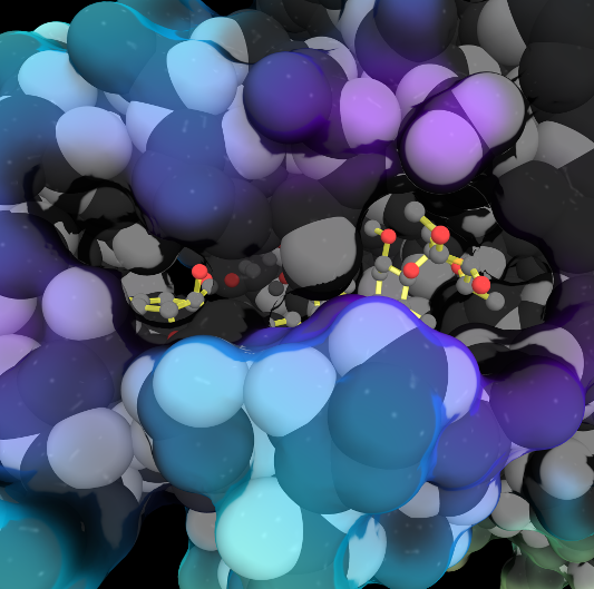
The X-ray structure of HIV-1 protease reveals that it is composed of two symmetrically related subunits which form a tunnel where they meet. This is critical because it contains the active site of the protease, consisting on two Asp-Thr-Gly conserved sequences, making it a member of the aspartyl protease family. The two catalytic Asp's either interact with the incoming water or protonate the carbonyl to make the carbon more electrophilic for the incoming water.

>>> Visit this page >>>

About this image
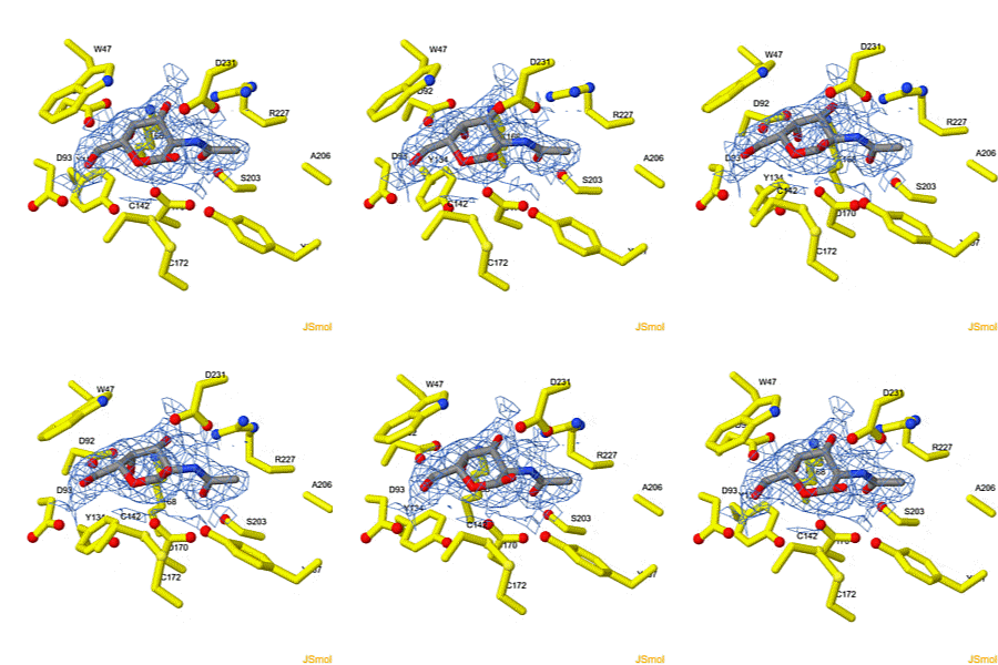
Molecular Sculpture

by Eric Martz
A historical review on sculptures and physical models of macromolecules.

>>> Visit this page >>>

About this image
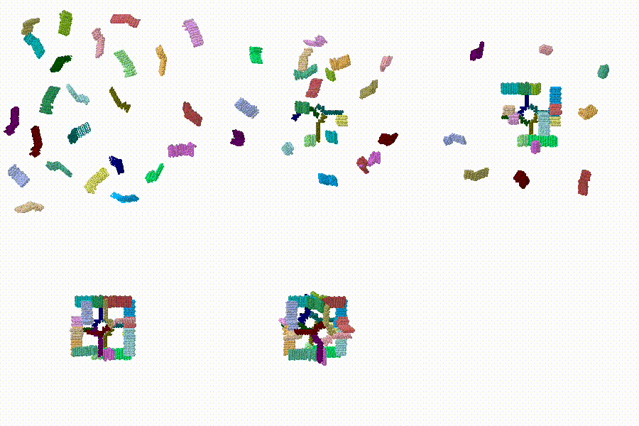
Structural flexibility of the periplasmic protein, FlgA, regulates flagellar P-ring assembly in Salmonella enterica.

H Matsunami, YH Yoon, VA Meshcheryakov, K Namba, FA Samatey. Scientific Reports 2016 doi: 10.1038/srep27399
A periplasmic flagellar chaperone protein, FlgA, is required for P-ring assembly in bacterial flagella of taxa such as Salmonella enterica or Escherichia coli. Here we present the open and closed crystal structures of FlgA from Salmonella enterica serovar Typhimurium, grown under different crystallization conditions. An intramolecular disulfide cross-linked form of FlgA caused a dominant negative effect on motility of the wild-type strain.

>>> Visit this I3DC complement >>>

About this image
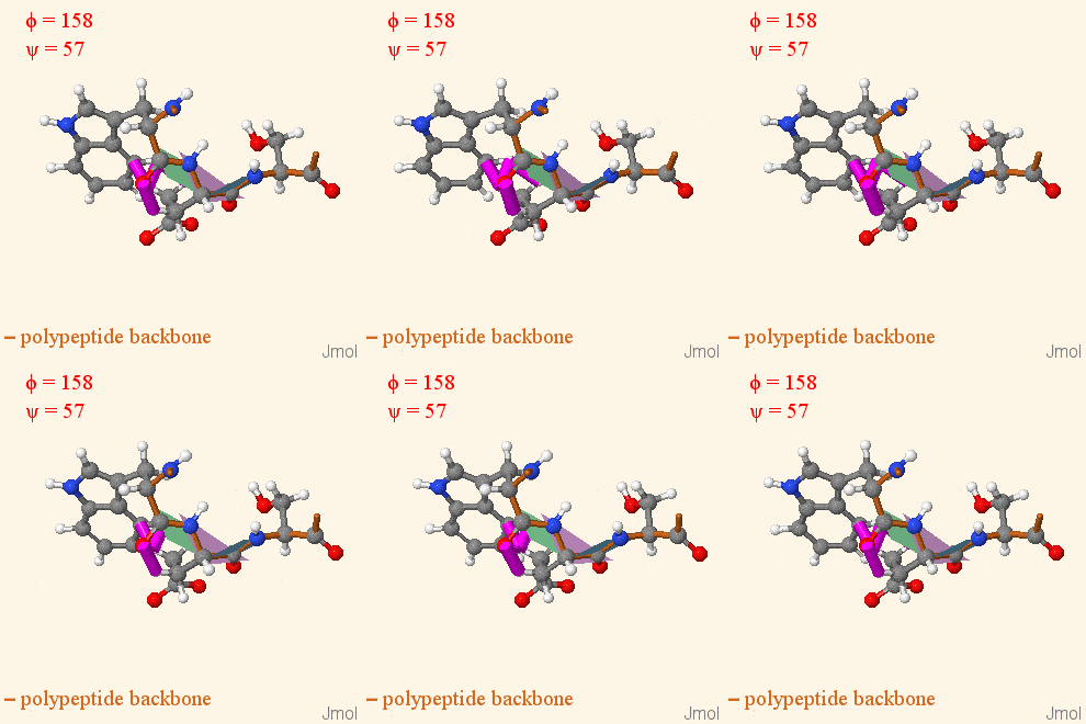
Make Your Own Electrostatic Potential Maps

Positive (+) and Negative (-) charges on the surface of a protein molecule play crucial roles in its interactions with other molecules, and hence in its functions. Electrostatic potential maps coloring the surface of a protein molecule are a popular way to visualize the distribution of surface charges. Easy to use free software is available to to create these surface maps. Above is an integral membrane potassium channel protein. One of its 4 identical chains is removed so you can see the Negative (-) protein surface contacting the 3 K+ ions.

>>> See Examples and Get Instructions >>>

How to add content to Proteopedia

Video Guides

Who knows ...

List of Proteopedia's Art on Science pages

About Interactive 3D Complements - I3DCs

List of I3DCs

How to get an I3DC for your paper

Teaching Strategies Using Proteopedia

Examples of Pages for Teaching

How to add content to Proteopedia

About Contact Table of Contents Structure Index Help

Proteopedia Page Contributors and Editors (what is this?)

Joel L. Sussman, Jaime Prilusky

Personal tools