Sandbox Reserved 1473
From Proteopedia
| Line 39: | Line 39: | ||
==STRUCTURE HIGHLIGHTS== | ==STRUCTURE HIGHLIGHTS== | ||
| - | + | ---- | |
| - | + | ||
'''Structural components of the B2 GPCR'''. | '''Structural components of the B2 GPCR'''. | ||
| - | + | ||
| + | <StructureSection load='2rh1' size='300' side='right' caption='Human Beta adrenergic receptor with bound ligand' scene='80/800652/Showing_ligand/1' > | ||
| + | |||
[[Image:Disulfide Linkages.png|thumb|downright=2.1| Disulfide Linkages between the TMD and ECL]] | [[Image:Disulfide Linkages.png|thumb|downright=2.1| Disulfide Linkages between the TMD and ECL]] | ||
Revision as of 01:44, 10 December 2018
| This Sandbox is Reserved from November 5 2018 through January 1, 2019 for use in the course "CHEM 4923: Senior Project taught by Christina R. Bourne at the University of Oklahoma, Norman, USA. This reservation includes Sandbox Reserved 1471 through Sandbox Reserved 1478. |
To get started:
More help: Help:Editing |
This page is reserved for Manny
Your Heading Here (maybe something like 'Structure')
| |||||||||||
References
- ↑ Hanson, R. M., Prilusky, J., Renjian, Z., Nakane, T. and Sussman, J. L. (2013), JSmol and the Next-Generation Web-Based Representation of 3D Molecular Structure as Applied to Proteopedia. Isr. J. Chem., 53:207-216. doi:http://dx.doi.org/10.1002/ijch.201300024
- ↑ Herraez A. Biomolecules in the computer: Jmol to the rescue. Biochem Mol Biol Educ. 2006 Jul;34(4):255-61. doi: 10.1002/bmb.2006.494034042644. PMID:21638687 doi:10.1002/bmb.2006.494034042644
THE GENUIS OF GPCRs.
|
NTRODUCTION.
Within the course of our daily activities, we experience several situations that cause us to jump on our feet and also, initiate some kind of response to these situations. Our bodies have an outstanding way of preparing us for such events as to whether flee or fight and it does this by making energy in the form of glucose available in the blood to support our reactions to these events. Our response is effectively mediated through reflex actions which are propagated through networks of neurons that are wired all over our body and eventually to the brain. One of the neurotransmitters produced through the coordination of these neuronal networks is epinephrine. Epinephrine serves as a ligand that bind to a transmembrane protein called GPCR and initiates a sequence of cascading pathways further downstream, that eventually results in the release of glucose into the blood to support the response we carry out.
These ligands bind to a receptor in the Transmembrane called GPCRs. Once the ligand is able to bind tightly to the receptor, the first committed step of the pathway is initiated and the connected pieces begin to join in together until blood glucose level is replenished. More than 800 genes code for GPCRs that regulate several signaling pathways which control blood pressure, behavior, sleep, immune response, cognition…and many other cellular processes.
STRUCTURE HIGHLIGHTS
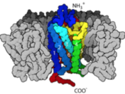
Structural components of the B2 GPCR.
| |||||||||||


