We apologize for Proteopedia being slow to respond. For the past two years, a new implementation of Proteopedia has been being built. Soon, it will replace this 18-year old system. All existing content will be moved to the new system at a date that will be announced here.
Sandbox Reserved 1473
From Proteopedia
(Difference between revisions)
| Line 3: | Line 3: | ||
This page is reserved for Manny | This page is reserved for Manny | ||
| - | == | + | ==THE GENUIS OF GPCRs == |
| + | <StructureSection load='2rh1' size='300' side='right' caption='Human Beta adrenergic receptor with bound ligand' scene='80/800652/Showing_ligand/1' > | ||
<StructureSection load='1stp' size='440' side='right' caption='Caption for this structure' scene=''> | <StructureSection load='1stp' size='440' side='right' caption='Caption for this structure' scene=''> | ||
| - | This is a default text for your page ''''''. Click above on '''edit this page''' to modify. Be careful with the < and > signs. | ||
You may include any references to papers as in: the use of JSmol in Proteopedia <ref>DOI 10.1002/ijch.201300024</ref> or to the article describing Jmol <ref>PMID:21638687</ref> to the rescue. | You may include any references to papers as in: the use of JSmol in Proteopedia <ref>DOI 10.1002/ijch.201300024</ref> or to the article describing Jmol <ref>PMID:21638687</ref> to the rescue. | ||
| - | == | + | == INTRODUCTION == |
| - | + | ||
| - | + | ||
| - | + | ||
| - | + | ||
| - | + | ||
| - | + | ||
| - | + | ||
| - | + | ||
| - | + | ||
| - | + | ||
| - | + | ||
| - | + | ||
| - | + | ||
| - | + | ||
| - | + | ||
| - | + | ||
| - | + | ||
| - | + | ||
| - | + | ||
| - | + | ||
| - | + | ||
| - | + | ||
Within the course of our daily activities, we experience several situations that cause us to jump on our feet and also, initiate some kind of response to these situations. Our bodies have an outstanding way of preparing us for such events as to whether flee or fight and it does this by making energy in the form of glucose available in the blood to support our reactions to these events. Our response is effectively mediated through reflex actions which are propagated through networks of neurons that are wired all over our body and eventually to the brain. One of the neurotransmitters produced through the coordination of these neuronal networks is epinephrine. Epinephrine serves as a ligand that bind to a transmembrane protein called GPCR and initiates a sequence of cascading pathways further downstream, that eventually results in the release of glucose into the blood to support the response we carry out. | Within the course of our daily activities, we experience several situations that cause us to jump on our feet and also, initiate some kind of response to these situations. Our bodies have an outstanding way of preparing us for such events as to whether flee or fight and it does this by making energy in the form of glucose available in the blood to support our reactions to these events. Our response is effectively mediated through reflex actions which are propagated through networks of neurons that are wired all over our body and eventually to the brain. One of the neurotransmitters produced through the coordination of these neuronal networks is epinephrine. Epinephrine serves as a ligand that bind to a transmembrane protein called GPCR and initiates a sequence of cascading pathways further downstream, that eventually results in the release of glucose into the blood to support the response we carry out. | ||
These ligands bind to a receptor in the Transmembrane called GPCRs. Once the ligand is able to bind tightly to the receptor, the first committed step of the pathway is initiated and the connected pieces begin to join in together until blood glucose level is replenished. More than 800 genes code for GPCRs that regulate several signaling pathways which control blood pressure, behavior, sleep, immune response, cognition…and many other cellular processes. | These ligands bind to a receptor in the Transmembrane called GPCRs. Once the ligand is able to bind tightly to the receptor, the first committed step of the pathway is initiated and the connected pieces begin to join in together until blood glucose level is replenished. More than 800 genes code for GPCRs that regulate several signaling pathways which control blood pressure, behavior, sleep, immune response, cognition…and many other cellular processes. | ||
| - | |||
| - | |||
| Line 42: | Line 18: | ||
'''Structural components of the B2 GPCR'''. | '''Structural components of the B2 GPCR'''. | ||
| - | |||
[[Image:Disulfide Linkages.png|thumb|downright=2.1| Disulfide Linkages between the TMD and ECL]] | [[Image:Disulfide Linkages.png|thumb|downright=2.1| Disulfide Linkages between the TMD and ECL]] | ||
| Line 49: | Line 24: | ||
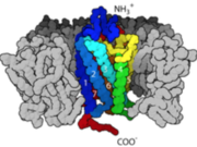
[[Image:Terminal.png|thumb|upleft=2.0| Amino and Carboxyl terminal groups]] | [[Image:Terminal.png|thumb|upleft=2.0| Amino and Carboxyl terminal groups]] | ||
| - | |||
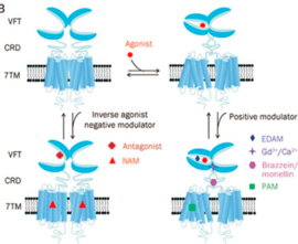
G-Protein Coupled Receptors (GPCR), also known as 7 transmembrane receptors [because of its 7 constituent alpha helices] are the largest groups of transmembrane protein receptors in eukaryotes. There are many different classes GPCRs depending on the position of their binding sites or the kind of ligands they bind to. There are about six classes of GPCRs and they include the classes A, B, C, D, E, AND F. Beta adrenergic receptors fall the class C with other metabotropic hormones while Rhodopsin (the light sensing cells in our eye) are categorized under class A. All studied GPCRs have a conserved structure that can be subdivided into; the extracellular domain (ECD), which comprises the three extracellular loops (EC1), (EC2), (EC3) and the N-terminal amino groups, the seven spanning helices transmembrane domain (7TM) embedded within the plasma membrane, and the intracellular domains ICD which also comprises the carboxyl-terminal residues and three intracellular loops (ICL1, ICL2, ICL3). The binding of the receptor to a ligand causes a structural change in the intracellular domains that also signals/activates the alpha subunit of a G-protein to exchange GDP for GTP, transducing the signal further downstream. | G-Protein Coupled Receptors (GPCR), also known as 7 transmembrane receptors [because of its 7 constituent alpha helices] are the largest groups of transmembrane protein receptors in eukaryotes. There are many different classes GPCRs depending on the position of their binding sites or the kind of ligands they bind to. There are about six classes of GPCRs and they include the classes A, B, C, D, E, AND F. Beta adrenergic receptors fall the class C with other metabotropic hormones while Rhodopsin (the light sensing cells in our eye) are categorized under class A. All studied GPCRs have a conserved structure that can be subdivided into; the extracellular domain (ECD), which comprises the three extracellular loops (EC1), (EC2), (EC3) and the N-terminal amino groups, the seven spanning helices transmembrane domain (7TM) embedded within the plasma membrane, and the intracellular domains ICD which also comprises the carboxyl-terminal residues and three intracellular loops (ICL1, ICL2, ICL3). The binding of the receptor to a ligand causes a structural change in the intracellular domains that also signals/activates the alpha subunit of a G-protein to exchange GDP for GTP, transducing the signal further downstream. | ||
| - | |||
For some GPCRs, the ligand binds to a site in the 7TM (certain class A) or both the ECD and the 7TM as in certain class B GPCRs. However, in the Beta Adrenergic receptor, the ligand binds at the the extracellular domain [ECD] which acts like a fly-trap pocket. The transmembrane domain is structurally coupled to the extracellular domain through Disulfide linkages and mutations within this region disrupts the entire structure and function of GPCRs. | For some GPCRs, the ligand binds to a site in the 7TM (certain class A) or both the ECD and the 7TM as in certain class B GPCRs. However, in the Beta Adrenergic receptor, the ligand binds at the the extracellular domain [ECD] which acts like a fly-trap pocket. The transmembrane domain is structurally coupled to the extracellular domain through Disulfide linkages and mutations within this region disrupts the entire structure and function of GPCRs. | ||
| Line 58: | Line 31: | ||
| - | + | == FUNCTION == | |
| - | + | ||
| - | == | + | |
GPCRs act as a bridge and communicate the conditions of the cell to the nucleus to induce transcription or affect the processivity of catalytic pathway by activating certain enzymes. In the event of fight/flight, upon binding the ligand epinephrine, it causes a conformational change that leads to the exchange of the Guanine nucleotide (GDP to GTP) and that mechanism "turns" the switch on. The alpha subunit then dissociate from the beta and gamma subunits and activates adenylate cyclase which makes cAMP. The cAMP activates Protein Kinase A by binding to its regulatory subunits, and that releases the catalytic subunits to further phosphorylate other proteins in the cell. Eventually, Glycogen phosphorylase is activated which breaks down glucose for use within the cell. | GPCRs act as a bridge and communicate the conditions of the cell to the nucleus to induce transcription or affect the processivity of catalytic pathway by activating certain enzymes. In the event of fight/flight, upon binding the ligand epinephrine, it causes a conformational change that leads to the exchange of the Guanine nucleotide (GDP to GTP) and that mechanism "turns" the switch on. The alpha subunit then dissociate from the beta and gamma subunits and activates adenylate cyclase which makes cAMP. The cAMP activates Protein Kinase A by binding to its regulatory subunits, and that releases the catalytic subunits to further phosphorylate other proteins in the cell. Eventually, Glycogen phosphorylase is activated which breaks down glucose for use within the cell. | ||
| Line 67: | Line 38: | ||
| + | == DISEASE == | ||
| - | ' | + | Many GPCRs have become drug targets of many pharmaceutical drugs because of their central role in controlling many metabolic activities in the cell. Diseases including cancer, diabetes, Alzheimer's, sleep deprivation and many others are treated by exploiting the druggability of GPCRs and how they mediate their responses to hormones, cytokines, and neurotransmitters. Of all drugs on the market that have been |
| + | approved by the CDC, about 35% elicit their potency and mediate their effects through GPCRs. | ||
| + | |||
| + | |||
| + | |||
| + | == ENERGETICS == | ||
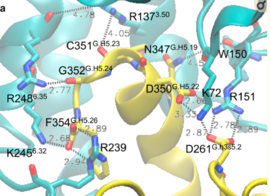
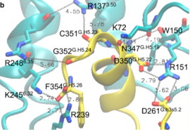
[[Image:Energetics1.png|thumb|upright=1.5| Salt bridge formation]] | [[Image:Energetics1.png|thumb|upright=1.5| Salt bridge formation]] | ||
| Line 81: | Line 58: | ||
| - | + | This is a sample scene created with SAT to <scene name="/12/3456/Sample/1">color</scene> by Group, and another to make <scene name="/12/3456/Sample/2">a transparent representation</scene> of the protein. You can make your own scenes on SAT starting from scratch or loading and editing one of these sample scenes. | |
| - | + | ||
| - | + | ||
| - | + | ||
| - | + | ||
| - | + | ||
| + | </StructureSection> | ||
== References == | == References == | ||
<references/> | <references/> | ||
Revision as of 19:02, 10 December 2018
| This Sandbox is Reserved from November 5 2018 through January 1, 2019 for use in the course "CHEM 4923: Senior Project taught by Christina R. Bourne at the University of Oklahoma, Norman, USA. This reservation includes Sandbox Reserved 1471 through Sandbox Reserved 1478. |
To get started:
More help: Help:Editing |
This page is reserved for Manny
THE GENUIS OF GPCRs
| |||||||||||
References
- ↑ Hanson, R. M., Prilusky, J., Renjian, Z., Nakane, T. and Sussman, J. L. (2013), JSmol and the Next-Generation Web-Based Representation of 3D Molecular Structure as Applied to Proteopedia. Isr. J. Chem., 53:207-216. doi:http://dx.doi.org/10.1002/ijch.201300024
- ↑ Herraez A. Biomolecules in the computer: Jmol to the rescue. Biochem Mol Biol Educ. 2006 Jul;34(4):255-61. doi: 10.1002/bmb.2006.494034042644. PMID:21638687 doi:10.1002/bmb.2006.494034042644
- ↑ Dong SS, Goddard WA 3rd, Abrol R. Conformational and Thermodynamic Landscape of GPCR Activation from Theory and Computation. Biophys J. 2016 Jun 21;110(12):2618-2629. doi: 10.1016/j.bpj.2016.04.028. PMID:27332120 doi:http://dx.doi.org/10.1016/j.bpj.2016.04.028


