Sandbox Reserved 1480
From Proteopedia
| Line 41: | Line 41: | ||
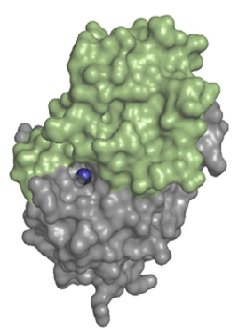
LHPP forms a homodimer in solution. Each <scene name='80/802654/2x4d_monomer/5'>monomer</scene> of hLHPP has two domains, one catalytic domain with a <scene name='80/802654/Mg_ion/2'>phosphoric acid and a magnesium ion</scene> (Mg2+) on the <scene name='80/802654/Catalityc_site/3'>active site</scene> and a large C2a-type cap domain which control the accessibility to the active site. Despite the large cap, the enzyme is not only able to act on small metabolites, but also on phosphoproteins. | LHPP forms a homodimer in solution. Each <scene name='80/802654/2x4d_monomer/5'>monomer</scene> of hLHPP has two domains, one catalytic domain with a <scene name='80/802654/Mg_ion/2'>phosphoric acid and a magnesium ion</scene> (Mg2+) on the <scene name='80/802654/Catalityc_site/3'>active site</scene> and a large C2a-type cap domain which control the accessibility to the active site. Despite the large cap, the enzyme is not only able to act on small metabolites, but also on phosphoproteins. | ||
| + | |||
The <scene name='80/802654/Catalityc_site_aa/3'>amino acids</scene> involved in the catalytic center are number 18 to 22, from 55 to 60, 155, 190, from 214 to 216, 219 and 220 corresponding respectively to DISGV, TNESAA, G, K, GDD, G and D. | The <scene name='80/802654/Catalityc_site_aa/3'>amino acids</scene> involved in the catalytic center are number 18 to 22, from 55 to 60, 155, 190, from 214 to 216, 219 and 220 corresponding respectively to DISGV, TNESAA, G, K, GDD, G and D. | ||
| + | [[Image:2 domains.png]] | ||
| + | |||
| + | ''Fig 4. Structure of a monomer of hLHPP. The catalytic domains is shown in gray, and the C2a-type cap domains is colored in green. The essential Mg2+ cofactor in the active site is shown as a blue sphere. Source : Do metabolic HAD phosphatases moonlight as protein phosphatases? Antje Gohla. (2018) [BBA - Molecular Cell Research]'' | ||
Revision as of 15:10, 10 January 2019
| This Sandbox is Reserved from 06/12/2018, through 30/06/2019 for use in the course "Structural Biology" taught by Bruno Kieffer at the University of Strasbourg, ESBS. This reservation includes Sandbox Reserved 1480 through Sandbox Reserved 1543. |
To get started:
More help: Help:Editing |
Structure of the protein LHPP
| |||||||||||
References
"Molecular cloning of a cDNA for the human phospholysine phosphohistidine inorganic pyrophosphate phosphatase." Yokoi F., Hiraishi H., Izuhara K. J. Biochem. 133:607-614(2003) [PubMed] [Europe PMC] [Abstract]
"The protein histidine phosphatase LHPP is a tumour suppressor." Sravanth K. Hindupur, Marco Colombi, Stephen R. Fuhs, Matthias S. Matter, Yakir Guri, Kevin Adam, Marion Cornu, Salvatore Piscuoglio, Charlotte K. Y. Ng, Charles Betz, Dritan Liko, Luca Quagliata, Suzette Moes, Paul Jenoe, Luigi M. Terracciano, Markus H. Heim, Tony Hunter & Michael N. Hall. (2018) [PubMed] [Main]
"Do metabolic HAD phosphatases moonlight as protein phosphatases?." Antje Gohla. (2018) [BBA - Molecular Cell Research]
"Down-regulation of LHPP in cervical cancer influences cell proliferation, metastasis and apoptosis by modulating AKT", (2018), [PubMed] [Main]


