Sandbox Reserved 1500
From Proteopedia
| Line 4: | Line 4: | ||
<table><tr><td colspan='2'>[[5hfg]] is a 1 chain structure. Full crystallographic information is available from [http://oca.weizmann.ac.il/oca-bin/ocashort?id=5HFG OCA]. For a <b>guided tour on the structure components</b> use [http://oca.weizmann.ac.il/oca-docs/fgij/fg.htm?mol=5HFG FirstGlance]. <br> | <table><tr><td colspan='2'>[[5hfg]] is a 1 chain structure. Full crystallographic information is available from [http://oca.weizmann.ac.il/oca-bin/ocashort?id=5HFG OCA]. For a <b>guided tour on the structure components</b> use [http://oca.weizmann.ac.il/oca-docs/fgij/fg.htm?mol=5HFG FirstGlance]. <br> | ||
</td></tr><tr id='related'><td class="sblockLbl"><b>[[Related_structure|Related:]]</b></td><td class="sblockDat">[[5hfi|5hfi]]</td></tr> | </td></tr><tr id='related'><td class="sblockLbl"><b>[[Related_structure|Related:]]</b></td><td class="sblockDat">[[5hfi|5hfi]]</td></tr> | ||
| - | </td></tr></td></tr><tr id='ligand'><td class="sblockLbl"><b>[[Ligand|Secondary structure:]]</b></td><td class="sblockDat"><scene name='pdbligand=ACT:ACETATE+ION'> | + | </td></tr></td></tr><tr id='ligand'><td class="sblockLbl"><b>[[Ligand|Secondary structure:]]</b></td><td class="sblockDat"><scene name='pdbligand=ACT:ACETATE+ION'>Beta sheets</scene>[[Image:5hfgplan.PNG|thumb|middle|'''Figure 1:''' Acetate ion.<ref>PDB image</ref>]], <scene name='pdbligand=CU:COPPER+(II)+ION'>alpha helix</scene></td></tr> |
<tr id='resources'><td class="sblockLbl"><b>Resources:</b></td><td class="sblockDat"><span class='plainlinks'>[http://oca.weizmann.ac.il/oca-docs/fgij/fg.htm?mol=5hfg FirstGlance], [http://oca.weizmann.ac.il/oca-bin/ocaids?id=5hfg OCA], [http://pdbe.org/5hfg PDBe], [http://www.rcsb.org/pdb/explore.do?structureId=5hfg RCSB], [http://www.ebi.ac.uk/pdbsum/5hfg PDBsum], [http://prosat.h-its.org/prosat/prosatexe?pdbcode=5hfg ProSAT]</span></td></tr> | <tr id='resources'><td class="sblockLbl"><b>Resources:</b></td><td class="sblockDat"><span class='plainlinks'>[http://oca.weizmann.ac.il/oca-docs/fgij/fg.htm?mol=5hfg FirstGlance], [http://oca.weizmann.ac.il/oca-bin/ocaids?id=5hfg OCA], [http://pdbe.org/5hfg PDBe], [http://www.rcsb.org/pdb/explore.do?structureId=5hfg RCSB], [http://www.ebi.ac.uk/pdbsum/5hfg PDBsum], [http://prosat.h-its.org/prosat/prosatexe?pdbcode=5hfg ProSAT]</span></td></tr> | ||
</table> | </table> | ||
Revision as of 21:53, 10 January 2019
| This Sandbox is Reserved from 06/12/2018, through 30/06/2019 for use in the course "Structural Biology" taught by Bruno Kieffer at the University of Strasbourg, ESBS. This reservation includes Sandbox Reserved 1480 through Sandbox Reserved 1543. |
To get started:
More help: Help:Editing |
Contents |
Structural highlights
| 5hfg is a 1 chain structure. Full crystallographic information is available from OCA. For a guided tour on the structure components use FirstGlance. | |
| Related: | 5hfi |
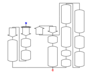
| Secondary structure: |  Figure 1: Acetate ion.[1] |
| Resources: | FirstGlance, OCA, PDBe, RCSB, PDBsum, ProSAT |
Global Symmetry: Asymmetric - C1
Global Stoichiometry: Monomer - A
Its theoretical weight is 25.29 KDa
Primary structure
5hfg is a 1 chain structure of 238 amino acids. [2]
Secondary structure
The structure of 5hfg mainly consists in alpha helix (12) , beta sheets (4), you can check the 3D view here. 
Tertiary structure
Monomeric assembly composition. 5hfg is one distinct polypeptide molecule. [3]
|
Nature
5hfg is a Dsb-family protein, from Pseudomonas Aeruginosa. This family of proteins is mainly used to oxidize and reduce cysteine residues of substrate proteins. Most enzymes from Dsb-family catalyze disulfide formation in periplasmic or secreted substrate proteins. [4] It has no bound ligands and no modified residues. Sequence domains:
   Thioredoxin-like superfamily
   DSBA-like thioredoxin domain
Function
This protein has a disulfide oxidoreductase activity.
Disease
Relevance
This is a sample scene created with SAT to by Group, and another to make of the protein.
</StructureSection>
