Main Page

From Proteopedia

(Difference between revisions)
Jump to: navigation, search
Line 8: Line 8:
<span style="border:none; margin:0; padding:0.3em; color:#000; font-style: italic; font-size: 1.1em;max-width:80%;display:block;">
-
Proteopedia's goal is to present structure/function information on 3D biomacromolecules in a user-friendly manner to a broad scientific audience.
+
Its goal is to present this information in a user-friendly manner to a broad scientific audience as a free, collaborative 3D-encyclopedia of proteins & other molecules.
</span>
</span>

Revision as of 09:05, 20 January 2019

ISSN 2310-6301

As life is more than 2D, Proteopedia helps to bridge the gap between 3D structure & function of biomacromolecules

Its goal is to present this information in a user-friendly manner to a broad scientific audience as a free, collaborative 3D-encyclopedia of proteins & other molecules.


Selected Pages Journals Education
About this image
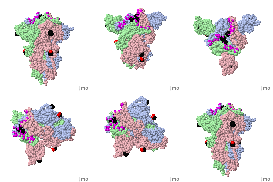
Green Fluorescent Protein

by Eran Hodis
Green fluorescent protein (GFP) is a bioluminescent polypeptide isolated from the jellyfish Aequorea victoria. GFP converts the blue chemiluminescence of aequorin into green fluorescent light. In the laboratory, GFP can be incorporated into a variety of biological systems in order to function as a marker protein. Since its discovery in 1962, GFP has become a significant contributor to the research of monitoring gene expression, localization, mobility, traffic, or interactions between various membrane and cytoplasmic proteins.

>>> Visit this page >>>

About this image
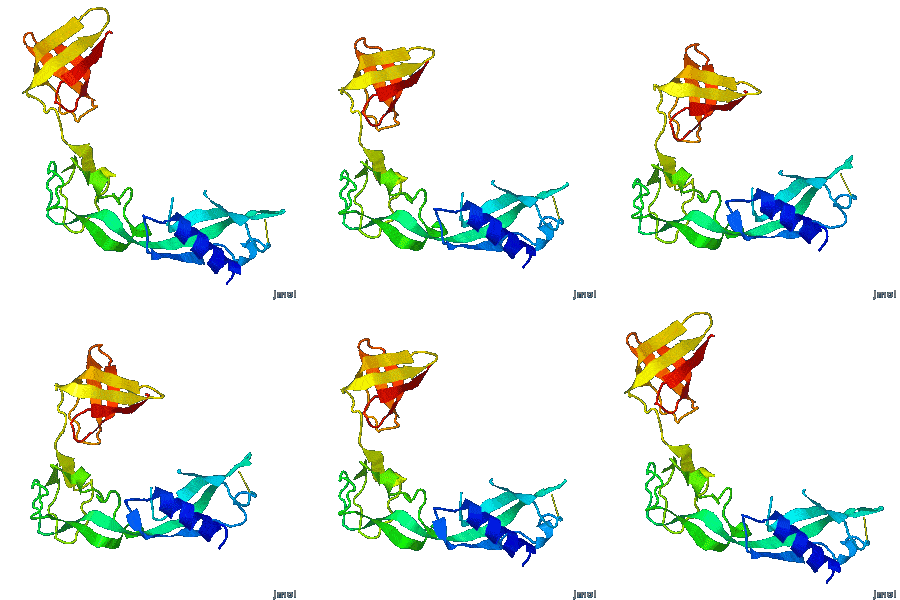
Structure of Anticancer Ruthenium Half-Sandwich Complex Bound to Glycogen Synthase Kinase 3ß

G Atilla-Gocumen, L Di Costanzo, E Meggers. J Biol Inorg Chem. 2010 doi: 10.1007/s00775-010-0699-x
A crystal structure of an organometallic half-sandwich ruthenium complex bound to glycogen synthase kinase 3ß (GSK-3ß) reveals that the inhibitor binds to the ATP binding site via an induced fit mechanism utilizing several hydrogen bonds and hydrophobic interactions. Importantly, the metal is not involved in any direct interaction with the protein kinase but fulfills a purely structural role.

>>> Visit this I3DC complement >>>

About this image
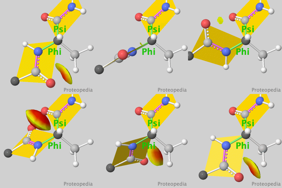
Tutorial: The Ramachandran principle, phi (φ) and psi (ψ) angles in proteins

by Eric Martz
The Ramachandran Principle says that alpha helices, beta strands, and turns are the most likely conformations for a polypeptide chain to adopt, because most other conformations are impossible due to steric collisions between atoms. Check Show Clashes to see where non-bonded atoms are overlapping, and thus in physically impossible positions.

>>> Visit this tutorial >>>

How to add content to Proteopedia

Video Guides

Who knows ...

About Interactive 3D Complements - I3DCs

List of I3DCs

How to get an I3DC for your paper

Teaching strategies using Proteopedia

Examples of pages for teaching

How to add content to Proteopedia

About Contact Table of Contents Structure Index Help

Proteopedia Page Contributors and Editors (what is this?)

Joel L. Sussman, Jaime Prilusky

Personal tools