Diphtheria toxin
From Proteopedia
(Difference between revisions)
| Line 15: | Line 15: | ||
== Structural highlights == | == Structural highlights == | ||
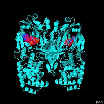
| - | <scene name='59/592684/Cv/ | + | <scene name='59/592684/Cv/17'>The biological assembly of Diphtheria toxin is dimer</scene>. DT is proteolitically cleaved into 2 fragments. <scene name='59/592684/Cv/7'>Fragment A contains the catalytic domain (C)</scene> and <scene name='59/592684/Cv/8'>fragment B</scene> contains the <scene name='59/592684/Cv/9'>transmembrane (T)</scene> and <scene name='59/592684/Cv/10'>receptor-binding (R)</scene> domains. <scene name='59/592684/Cv/15'>DT active site</scene> is located in a cleft in the C domain.<ref>PMID:7833807</ref> Water molecules are shown as red spheres. The 2 monomers of DT interact by domain swapping to form a <scene name='59/592684/Cv/16'>compact, globular dimer structure</scene>. |
</StructureSection> | </StructureSection> | ||
Revision as of 10:47, 24 February 2019
| |||||||||||
3D structures of diphtheria toxin
Updated on 24-February-2019
References
- ↑ Pappenheimer AM Jr. Diphtheria toxin. Annu Rev Biochem. 1977;46:69-94. PMID:20040 doi:http://dx.doi.org/10.1146/annurev.bi.46.070177.000441
- ↑ Bennett MJ, Choe S, Eisenberg D. Refined structure of dimeric diphtheria toxin at 2.0 A resolution. Protein Sci. 1994 Sep;3(9):1444-63. PMID:7833807
