This old version of Proteopedia is provided for student assignments while the new version is undergoing repairs. Content and edits done in this old version of Proteopedia after March 1, 2026 will eventually be lost when it is retired in about June of 2026.
Apply for new accounts at the new Proteopedia. Your logins will work in both the old and new versions.
Adhesin
From Proteopedia
(Difference between revisions)
| Line 14: | Line 14: | ||
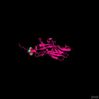
*<scene name='70/705684/Cv/3'>Galactose binding site</scene>. Water molecules are shown as red spheres. | *<scene name='70/705684/Cv/3'>Galactose binding site</scene>. Water molecules are shown as red spheres. | ||
*<scene name='70/705684/Cv/5'>Tert-butyl formate/Acetate binding site</scene>. | *<scene name='70/705684/Cv/5'>Tert-butyl formate/Acetate binding site</scene>. | ||
| + | |||
| + | == 3D Structures of adhesin == | ||
| + | [[Adhesin 3D structures]] | ||
| + | |||
</StructureSection> | </StructureSection> | ||
== 3D Structures of adhesin == | == 3D Structures of adhesin == | ||
| Line 28: | Line 32: | ||
**[[4b4p]], [[4bwo]] – EcF18 lectin domain <BR /> | **[[4b4p]], [[4bwo]] – EcF18 lectin domain <BR /> | ||
**[[4auu]], [[1uwf]], [[1tr7]], [[5fwr]] – EcAdh FimH lectin domain <BR /> | **[[4auu]], [[1uwf]], [[1tr7]], [[5fwr]] – EcAdh FimH lectin domain <BR /> | ||
| - | **[[5mca]], [[5fx3]] – EcAdh FimH lectin domain (mutant) <BR /> | + | **[[5mca]], [[5fx3]], [[5fs5]] – EcAdh FimH lectin domain (mutant) <BR /> |
**[[5afo]] – EcAdh LF82 <BR /> | **[[5afo]] – EcAdh LF82 <BR /> | ||
**[[6aow]] – EcAdh FMLH lectin domain <BR /> | **[[6aow]] – EcAdh FMLH lectin domain <BR /> | ||
| Line 34: | Line 38: | ||
**[[3zk8]], [[3zk9]], [[3zka]] – SpPsaA (mutant)<br /> | **[[3zk8]], [[3zk9]], [[3zka]] – SpPsaA (mutant)<br /> | ||
**[[3u4k]] – Adh lectin domain – ''Klebsiella pneumoniae''<BR /> | **[[3u4k]] – Adh lectin domain – ''Klebsiella pneumoniae''<BR /> | ||
| + | **[[6h1q]] – Adh – ''Proteus mirabilis'' <br /> | ||
*Fimbrial adhesion complexes | *Fimbrial adhesion complexes | ||
| Line 40: | Line 45: | ||
**[[4w6w]], [[4w6x]], [[4w6y]] – EcF18 lectin domain + nanobody <BR /> | **[[4w6w]], [[4w6x]], [[4w6y]] – EcF18 lectin domain + nanobody <BR /> | ||
**[[4j3o]] – EcAdh FimH + FimG + FimC + FimF + FimD <BR /> | **[[4j3o]] – EcAdh FimH + FimG + FimC + FimF + FimD <BR /> | ||
| + | **[[6e14]], [[6e15]] – EcAdh FimH + FimG + FimC + FimF + FimD - Cryo EM<BR /> | ||
**[[3rfz]], [[1ze3]] – EcAdh FimH + FimC + FimD <BR /> | **[[3rfz]], [[1ze3]] – EcAdh FimH + FimC + FimD <BR /> | ||
**[[1qun]], [[1klf]], [[1kiu]] – EcAdh FimH + FimC <BR /> | **[[1qun]], [[1klf]], [[1kiu]] – EcAdh FimH + FimC <BR /> | ||
| Line 49: | Line 55: | ||
**[[6aox]] – EcAdh FMLD lectin domain + TF antigen <BR /> | **[[6aox]] – EcAdh FMLD lectin domain + TF antigen <BR /> | ||
**[[6aro]] – EcAdh FMLH lectin domain + quinoline derivative <BR /> | **[[6aro]] – EcAdh FMLH lectin domain + quinoline derivative <BR /> | ||
| - | **[[5muc]], [[5l4y]], [[5l4x]], [[5l4w]], [[5l4v]], [[5l4u]], [[5l4t]], [[5jcq]], [[5jcr]], [[5f2f]], [[5f3f]], [[5abz]], [[4xo8]], [[4x5p]], [[4x5q]], [[4x5r]], [[4lov]], [[4css]], [[4cst]], [[4att]], [[4auj]], [[4auy]], [[4av0]], [[4av4]], [[4av5]], [[4avh]], [[4avi]], [[4avj]], [[4avk]], [[3zl1]], [[3zl2]], [[2vco]] – EcAdh FimH lectin domain + mannoside derivative <BR /> | + | **[[5muc]], [[5l4y]], [[5l4x]], [[5l4w]], [[5l4v]], [[5l4u]], [[5l4t]], [[5jcq]], [[5jcr]], [[5f2f]], [[5f3f]], [[5abz]], [[4xo8]], [[4x5p]], [[4x5q]], [[4x5r]], [[4lov]], [[4css]], [[4cst]], [[4att]], [[4auj]], [[4auy]], [[4av0]], [[4av4]], [[4av5]], [[4avh]], [[4avi]], [[4avj]], [[4avk]], [[3zl1]], [[3zl2]], [[2vco]], [[6gtx]], [[6gty]] – EcAdh FimH lectin domain + mannoside derivative <BR /> |
**[[5cgb]] – EcAdh FimH lectin domain + galactitol <BR /> | **[[5cgb]] – EcAdh FimH lectin domain + galactitol <BR /> | ||
**[[5ab1]], [[4x50]] – EcAdh FimH + mannoside derivative <BR /> | **[[5ab1]], [[4x50]] – EcAdh FimH + mannoside derivative <BR /> | ||
| Line 116: | Line 122: | ||
**[[3d9x]] – Adh A – ''Bartonella henselae'' <BR /> | **[[3d9x]] – Adh A – ''Bartonella henselae'' <BR /> | ||
**[[2lwb]] – Adh WI-1 – ''Ajellomyces dermatitidis'' <BR /> | **[[2lwb]] – Adh WI-1 – ''Ajellomyces dermatitidis'' <BR /> | ||
| - | **[[5my7]] – | + | **[[5my7]] – NmAdh residues 1-124 – ''Neisseria meningitidis'' <BR /> |
| + | **[[6eun]] – NmAdh residues 24-170<BR /> | ||
| + | **[[6eup]] – NmAdh residues 24-170 (mutant)<BR /> | ||
| + | **[[6gq4]] – Adh residues 83-185 – ''Neisseria gonorheae''<BR /> | ||
}} | }} | ||
Revision as of 09:33, 3 March 2019
| |||||||||||
3D Structures of adhesin
Updated on 03-March-2019
References
- ↑ Klemm P, Schembri MA. Bacterial adhesins: function and structure. Int J Med Microbiol. 2000 Mar;290(1):27-35. PMID:11043979 doi:http://dx.doi.org/10.1016/S1438-4221(00)80102-2
- ↑ Nishiyama M, Ishikawa T, Rechsteiner H, Glockshuber R. Reconstitution of pilus assembly reveals a bacterial outer membrane catalyst. Science. 2008 Apr 18;320(5874):376-9. doi: 10.1126/science.1154994. Epub 2008 Mar, 27. PMID:18369105 doi:http://dx.doi.org/10.1126/science.1154994
- ↑ Wizemann TM, Adamou JE, Langermann S. Adhesins as targets for vaccine development. Emerg Infect Dis. 1999 May-Jun;5(3):395-403. doi: 10.3201/eid0503.990310. PMID:10341176 doi:http://dx.doi.org/10.3201/eid0503.990310
- ↑ Bao R, Nair MK, Tang WK, Esser L, Sadhukhan A, Holland RL, Xia D, Schifferli DM. Structural basis for the specific recognition of dual receptors by the homopolymeric pH 6 antigen (Psa) fimbriae of Yersinia pestis. Proc Natl Acad Sci U S A. 2013 Jan 15;110(3):1065-70. doi:, 10.1073/pnas.1212431110. Epub 2012 Dec 31. PMID:23277582 doi:10.1073/pnas.1212431110
