We apologize for Proteopedia being slow to respond. For the past two years, a new implementation of Proteopedia has been being built. Soon, it will replace this 18-year old system. All existing content will be moved to the new system at a date that will be announced here.
Epoxide hydrolase
From Proteopedia
(Difference between revisions)
| Line 11: | Line 11: | ||
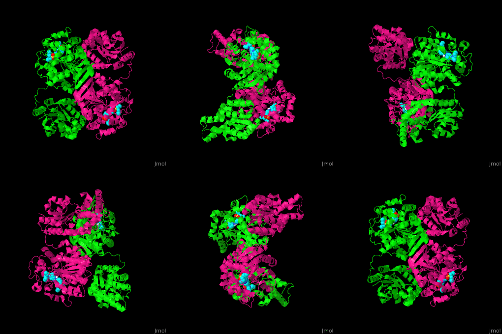
== Structural highlights == | == Structural highlights == | ||
| - | <scene name='70/708145/Cv/ | + | <scene name='70/708145/Cv/6'>Biological assembly of Human soluble epoxide hydrolase is dimer</scene>. |
| - | <scene name='70/708145/Cv/ | + | <scene name='70/708145/Cv/7'>Inhibitor binding site</scene> (PDB code [[5alf]]).<ref>PMID:25931264</ref> |
</StructureSection> | </StructureSection> | ||
Revision as of 10:04, 12 March 2019
| |||||||||||
3D Structures of epoxide hydrolase
Updated on 12-March-2019
References
- ↑ Biswal BK, Morisseau C, Garen G, Cherney MM, Garen C, Niu C, Hammock BD, James MN. The molecular structure of epoxide hydrolase B from Mycobacterium tuberculosis and its complex with a urea-based inhibitor. J Mol Biol. 2008 Sep 12;381(4):897-912. Epub 2008 Jun 17. PMID:18585390 doi:10.1016/j.jmb.2008.06.030
- ↑ Arand M, Hallberg BM, Zou J, Bergfors T, Oesch F, van der Werf MJ, de Bont JA, Jones TA, Mowbray SL. Structure of Rhodococcus erythropolis limonene-1,2-epoxide hydrolase reveals a novel active site. EMBO J. 2003 Jun 2;22(11):2583-92. PMID:12773375 doi:http://dx.doi.org/10.1093/emboj/cdg275
- ↑ Imig JD, Hammock BD. Soluble epoxide hydrolase as a therapeutic target for cardiovascular diseases. Nat Rev Drug Discov. 2009 Oct;8(10):794-805. doi: 10.1038/nrd2875. PMID:19794443 doi:http://dx.doi.org/10.1038/nrd2875
- ↑ Oster L, Tapani S, Xue Y, Kack H. Successful generation of structural information for fragment-based drug discovery. Drug Discov Today. 2015 Apr 28. pii: S1359-6446(15)00154-3. doi:, 10.1016/j.drudis.2015.04.005. PMID:25931264 doi:http://dx.doi.org/10.1016/j.drudis.2015.04.005

