User:Caitlin Marie Gaich/Sandbox1
From Proteopedia
(Difference between revisions)
| Line 3: | Line 3: | ||
=Histones= | =Histones= | ||
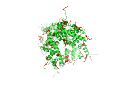
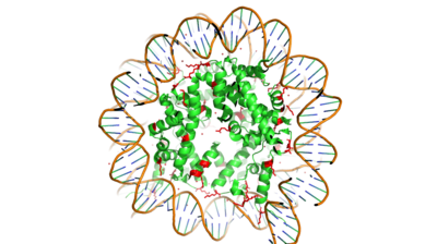
| - | [https://en.wikipedia.org/wiki/Histone Histones] are proteins found in the nucleus that are the key building blocks of [https://en.wikipedia.org/wiki/Chromatin chromatin] and are essential for proper DNA packaging and transcription. In the first step of DNA packaging, two copies of the four core histone proteins (H1A, H2A, H3, and H4) form an octomer in which DNA directly interacts with and wraps around, forming the nucleosome. 20-24% of residues making up the histone octomer are arginine and lysine, causing a net positive charge, especially at the outer surfaces of the histone core where negatively-charged DNA is bound. | + | [https://en.wikipedia.org/wiki/Histone Histones] are proteins found in the nucleus that are the key building blocks of [https://en.wikipedia.org/wiki/Chromatin chromatin] and are essential for proper DNA packaging and transcription. In the first step of DNA packaging, two copies of the four core histone proteins (H1A, H2A, H3, and H4) form an octomer in which DNA directly interacts with and wraps around, forming the nucleosome. 20-24% of residues making up the histone octomer are arginine and lysine, causing a net positive charge, especially at the outer surfaces of the histone core where negatively-charged DNA is bound. <ref> Watson, J D, et al. Molecular Biology of the Gene (Seventh Edition). (2014) Boston, MA: Benjamin-Cummings Publishing Company. </ref> It is those positively charged tails of the histone core that are often subject to post-translational modifications that play important roles in replication, transcription, heterochromatin maintenance, and DNA repair. |
| + | |||
| + | [[Image:Histone_Core.png|400 px|left|thumb|Figure 1. Histone Core-Lysine Residues Shown in Red]] | ||
| + | [[Image:Histone_w_DNA.png|400 px|right|thumb|Figure 2. Histone Core w/ DNA Bound]] | ||
| + | |||
==Histone Modification== | ==Histone Modification== | ||
| - | Histones can be modified in a variety of ways, including: | + | Histones can be modified in a variety of ways, including: methylation, demethylation, acetylation, deacetylation and many others, all leading to either the condensation or relaxation of DNA and as a consequence turning on or off DNA transcription. Histone acetylation is a common histone modification. This involves the transfer of an acetyl moiety from Acetyl Coenzyme A (AcCoA) to an ε-amino group of the target lysine residue on a histone. This reaction is catalyzed by the histone acetyltransferase (HAT) enzyme families. The specific histone acetylation modification is an important [https://en.wikipedia.org/wiki/Epigenetics epigenetic] marker. It plays a role in RNA synthesis and there a known correlation between gene activity and histone acetylation. Any misregulations of the HAT enzyme can possibly lead to cancer, cardiovascular disease, and HIV. |
=HAT1 Background = | =HAT1 Background = | ||
| Line 18: | Line 22: | ||
===Ligand Active Site=== | ===Ligand Active Site=== | ||
| - | [[Image:Binding interface.png|400 px|left|thumb|Figure | + | [[Image:Binding interface.png|400 px|left|thumb|Figure 3. Acetyl-CoA in the binding pocket of HAT1 (Chain A)]] |
The acetyl-CoA HAT1 active site is parallel to the C-terminal domain of the HAT1 protein. Acetyl-CoA fits structurally into the small binding site due to the kinked pantetheine group giving the molecule a bent confirmation. Once bound, most of the acetyl-CoA molecule is buried in the protein, around 60% (Figure 1). Hydrophobic contacts, hydrogen bonds, and salt bridges help to stabilize the protein-ligand interaction. HAT1 protein-ligand contact is concentrated in three areas: C-terminal end of helix alpha 7, C terminal end of strand beta-14/loop Beta15-Alpha9, and N-terminal half of helix alpha 9 <ref name=”Dutnall”>PMID:10384314</ref>. | The acetyl-CoA HAT1 active site is parallel to the C-terminal domain of the HAT1 protein. Acetyl-CoA fits structurally into the small binding site due to the kinked pantetheine group giving the molecule a bent confirmation. Once bound, most of the acetyl-CoA molecule is buried in the protein, around 60% (Figure 1). Hydrophobic contacts, hydrogen bonds, and salt bridges help to stabilize the protein-ligand interaction. HAT1 protein-ligand contact is concentrated in three areas: C-terminal end of helix alpha 7, C terminal end of strand beta-14/loop Beta15-Alpha9, and N-terminal half of helix alpha 9 <ref name=”Dutnall”>PMID:10384314</ref>. | ||
| Line 28: | Line 32: | ||
Of the five classes of HAT enzymes, the catalytic mechanisms for two of those enzymes, HAT1 and Rtt109, remains unclear. A structural overlay of HAT1 and Gcn5, a better-understood HAT enzyme, found a conserved glutamate residue in the active site of both molecules. Previous studies found that a mutation at the active site glutamate residue greatly alters the catalytic ability of HAT1, proving it to be structurally important. <ref> DOI:10.1101/gad.240531.114 </ref> Using this information and structural information from the crystallized structure of the HAT1/HAT2 complex regarding the proximity of potentially catalytic residues, the most plausible mechanism for histone acetylation involves the following relevant residues and cofactor: <scene name='81/811713/Mechanism_glu_lys_coa/1'>Glu255, Lys14, and AcetylCoA</scene>. | Of the five classes of HAT enzymes, the catalytic mechanisms for two of those enzymes, HAT1 and Rtt109, remains unclear. A structural overlay of HAT1 and Gcn5, a better-understood HAT enzyme, found a conserved glutamate residue in the active site of both molecules. Previous studies found that a mutation at the active site glutamate residue greatly alters the catalytic ability of HAT1, proving it to be structurally important. <ref> DOI:10.1101/gad.240531.114 </ref> Using this information and structural information from the crystallized structure of the HAT1/HAT2 complex regarding the proximity of potentially catalytic residues, the most plausible mechanism for histone acetylation involves the following relevant residues and cofactor: <scene name='81/811713/Mechanism_glu_lys_coa/1'>Glu255, Lys14, and AcetylCoA</scene>. | ||
| - | [[Image:HAT1_Mechanism.jpg|400px|right|thumb|Figure | + | [[Image:HAT1_Mechanism.jpg|400px|right|thumb|Figure 4: HAT1 Arrow-Pushing Mechanism]] |
In this mechanism, the glutamate at residue 255 in the active site of the protein acts as a general base by deprotonating lysine 12 of histone 4 (the numbering of the modified lysine residue on histone 4 is shifted two residues in the pdb file 4psw).The deprotonated lysine then acts as a nucleophile and attacks the carbonyl carbon of acetyl coenzyme Acetyl CoA (not shown in pdb file), forming a tetrahedral transition state with an oxyanion. The negative charge on the oxyanion then shift to down to reform the double bond between the oxygen and carbonyl carbon, breaking the scissle bond between the carbonyl carbon and the sulfur atom of acetyl CoA. The resulting product of this reaction is histone 4 with an acetyl-lysine at residue 12 and CoEnzyme A. | In this mechanism, the glutamate at residue 255 in the active site of the protein acts as a general base by deprotonating lysine 12 of histone 4 (the numbering of the modified lysine residue on histone 4 is shifted two residues in the pdb file 4psw).The deprotonated lysine then acts as a nucleophile and attacks the carbonyl carbon of acetyl coenzyme Acetyl CoA (not shown in pdb file), forming a tetrahedral transition state with an oxyanion. The negative charge on the oxyanion then shift to down to reform the double bond between the oxygen and carbonyl carbon, breaking the scissle bond between the carbonyl carbon and the sulfur atom of acetyl CoA. The resulting product of this reaction is histone 4 with an acetyl-lysine at residue 12 and CoEnzyme A. | ||
Revision as of 01:44, 13 April 2019
Histone Acetyltransferase HAT1/HAT2 Complex, Saccharomyces cerevisiae
| |||||||||||


