Diacylglycerol kinase
From Proteopedia
(Difference between revisions)
| Line 11: | Line 11: | ||
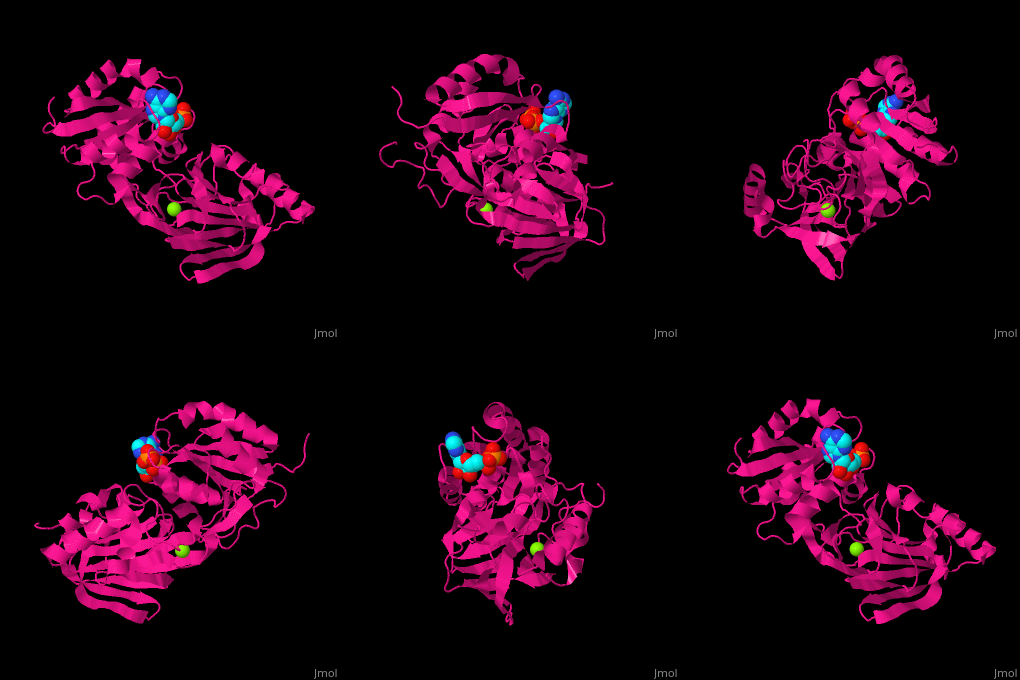
In DAGK DgkB the <scene name='70/706738/Cv/3'>nucleotide binding site</scene> is found at the interface between the 2 domains of the structure. <ref>PMID:18611377</ref> Water molrcules are shown asred spheres. | In DAGK DgkB the <scene name='70/706738/Cv/3'>nucleotide binding site</scene> is found at the interface between the 2 domains of the structure. <ref>PMID:18611377</ref> Water molrcules are shown asred spheres. | ||
| + | |||
| + | == 3D Structures of diacylglycerol kinase == | ||
| + | [[Diacylglycerol kinase 3D structures]] | ||
</StructureSection> | </StructureSection> | ||
| Line 21: | Line 24: | ||
*Diacylglycerol kinase DgkA | *Diacylglycerol kinase DgkA | ||
| + | **[[1tuz]] – hDAGK α residues 1-118 – human<br /> | ||
| + | **[[6iie]] – hDAGK α EF hand domain residues 107-197<br /> | ||
| + | **[[3bq7]] – hDAGK δ1 SAM domain (mutant) <br /> | ||
| + | **[[1r79]] – hDAGK δ1 C1 domain - NMR<br /> | ||
**[[3ze4]], [[4up6]], [[5d6i]], [[5dwk]] – EcDAGK – ''Escherichia coli'' <br /> | **[[3ze4]], [[4up6]], [[5d6i]], [[5dwk]] – EcDAGK – ''Escherichia coli'' <br /> | ||
**[[2kdc]] – EcDAGK - NMR <br /> | **[[2kdc]] – EcDAGK - NMR <br /> | ||
**[[3ze3]], [[3ze5]], [[4bpd]], [[4brb]], [[4brr]], [[4d2e]], [[4cjz]], [[4ck0]], [[4uxw]], [[4uxz]], [[4uyo]], [[5d56]], [[5d57]] – EcDAGK (mutant) <br /> | **[[3ze3]], [[3ze5]], [[4bpd]], [[4brb]], [[4brr]], [[4d2e]], [[4cjz]], [[4ck0]], [[4uxw]], [[4uxz]], [[4uyo]], [[5d56]], [[5d57]] – EcDAGK (mutant) <br /> | ||
**[[4uxx]] – EcDAGK (mutant) + AMPPCP<br /> | **[[4uxx]] – EcDAGK (mutant) + AMPPCP<br /> | ||
| - | **[[1tuz]] – hDAGK α – human<br /> | ||
| - | **[[3bq7]] – hDAGK δ1 SAM domain (mutant) <br /> | ||
| - | **[[1r79]] – hDAGK δ1 C1 domain - NMR<br /> | ||
**[[3s40]] – DAGK – Bacillus anthracis<br /> | **[[3s40]] – DAGK – Bacillus anthracis<br /> | ||
**[[4wer]] – DAGK catalytic domain – ''Enterococcus faecalis''<br /> | **[[4wer]] – DAGK catalytic domain – ''Enterococcus faecalis''<br /> | ||
| Line 33: | Line 37: | ||
*Diacylglycerol kinase DgkB | *Diacylglycerol kinase DgkB | ||
| - | **[[2qvl]] – | + | **[[2qvl]] – SaDgkB – ''Staphylococcus aureus''<br /> |
| - | **[[2qv7]] – | + | **[[2qv7]] – SaDgkB + ADP<br /> |
}} | }} | ||
== References == | == References == | ||
<references/> | <references/> | ||
[[Category:Topic Page]] | [[Category:Topic Page]] | ||
Revision as of 09:35, 5 June 2019
| |||||||||||
3D Structures of diacylglycerol kinase
Updated on 05-June-2019
References
- ↑ Merida I, Avila-Flores A, Merino E. Diacylglycerol kinases: at the hub of cell signalling. Biochem J. 2008 Jan 1;409(1):1-18. PMID:18062770 doi:http://dx.doi.org/10.1042/BJ20071040
- ↑ Miller DJ, Jerga A, Rock CO, White SW. Analysis of the Staphylococcus aureus DgkB structure reveals a common catalytic mechanism for the soluble diacylglycerol kinases. Structure. 2008 Jul;16(7):1036-46. PMID:18611377 doi:http://dx.doi.org/10.1016/j.str.2008.03.019

