We apologize for Proteopedia being slow to respond. For the past two years, a new implementation of Proteopedia has been being built. Soon, it will replace this 18-year old system. All existing content will be moved to the new system at a date that will be announced here.
NAD synthase
From Proteopedia
(Difference between revisions)
| Line 5: | Line 5: | ||
== Structural highlights == | == Structural highlights == | ||
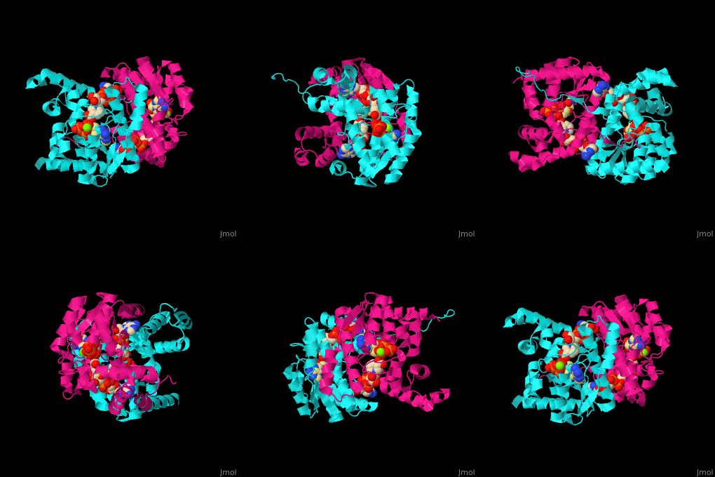
| - | The <scene name='54/541072/Cv/ | + | The <scene name='54/541072/Cv/8'>ATP binding site</scene> contains <scene name='54/541072/Cv/9'>Mg+2 ion</scene>. The <scene name='54/541072/Cv/10'>NAD binding site is located in the interface of the 2 subunits</scene><ref>PMID:15645437</ref>. Water molecules are shown as red spheres. |
</StructureSection> | </StructureSection> | ||
== 3D Structures of NAD+ synthase == | == 3D Structures of NAD+ synthase == | ||
Revision as of 12:15, 21 July 2019
| |||||||||||
3D Structures of NAD+ synthase
Updated on 21-July-2019
References
- ↑ Suda Y, Tachikawa H, Yokota A, Nakanishi H, Yamashita N, Miura Y, Takahashi N. Saccharomyces cerevisiae QNS1 codes for NAD(+) synthetase that is functionally conserved in mammals. Yeast. 2003 Aug;20(11):995-1005. PMID:12898714 doi:http://dx.doi.org/10.1002/yea.1008
- ↑ Kang GB, Kim YS, Im YJ, Rho SH, Lee JH, Eom SH. Crystal structure of NH3-dependent NAD+ synthetase from Helicobacter pylori. Proteins. 2005 Mar 1;58(4):985-8. PMID:15645437 doi:10.1002/prot.20377

