Protein kinase C
From Proteopedia
(Difference between revisions)
| Line 12: | Line 12: | ||
== Structural highlights == | == Structural highlights == | ||
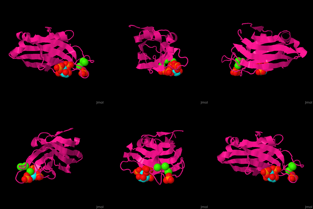
| - | <scene name='46/468129/Cv/ | + | <scene name='46/468129/Cv/6'>Phosphatidylinositol binds to PKC-α C2 domain groove</scene> near the <scene name='46/468129/Cv/7'>Ca+2 binding pocket</scene><ref>PMID:19346474</ref>. Water molecules are shown as red spheres. |
</StructureSection> | </StructureSection> | ||
==3D structures of protein kinase C== | ==3D structures of protein kinase C== | ||
Revision as of 14:08, 18 August 2019
| |||||||||||
3D structures of protein kinase C
Updated on 18-August-2019
References
- ↑ Idris I, Gray S, Donnelly R. Protein kinase C activation: isozyme-specific effects on metabolism and cardiovascular complications in diabetes. Diabetologia. 2001 Jun;44(6):659-73. PMID:11440359 doi:http://dx.doi.org/10.1007/s001250051675
- ↑ Koya D, King GL. Protein kinase C activation and the development of diabetic complications. Diabetes. 1998 Jun;47(6):859-66. PMID:9604860
- ↑ Koivunen J, Aaltonen V, Peltonen J. Protein kinase C (PKC) family in cancer progression. Cancer Lett. 2006 Apr 8;235(1):1-10. PMID:15907369 doi:http://dx.doi.org/10.1016/j.canlet.2005.03.033
- ↑ Guerrero-Valero M, Ferrer-Orta C, Querol-Audi J, Marin-Vicente C, Fita I, Gomez-Fernandez JC, Verdaguer N, Corbalan-Garcia S. Structural and mechanistic insights into the association of PKC{alpha}-C2 domain to PtdIns(4,5)P2. Proc Natl Acad Sci U S A. 2009 Apr 3. PMID:19346474
Proteopedia Page Contributors and Editors (what is this?)
Michal Harel, Alexander Berchansky, Joel L. Sussman, Jaime Prilusky

