Insulin-Degrading Enzyme
From Proteopedia
(Difference between revisions)
| Line 44: | Line 44: | ||
Virtual screening of the IDE protein (PDB ID: [[2jg4]]) was conducted using the binding site defined by the catalytic site of IDE protein. In silico results indicate that traditional Chinese medicine compounds <scene name='Journal:JBSD:29/Cv/5'>dihydrocaffeic acid</scene> (<span style="color:royalblue;background-color:black;font-weight:bold;">colored in royalblue</span>), <scene name='Journal:JBSD:29/Cv/6'>isopraeroside IV</scene> (<font color='blueviolet'><b>colored in blueviolet</b></font>), and <scene name='Journal:JBSD:29/Cv/8'>scopolin</scene> (<span style="color:chocolate;background-color:black;font-weight:bold;">colored in chocolate</span>) had high binding affinity with IDE protein and formed hydrogen bonds with the key active residue, <font color='magenta'><b>Glu111 (colored in magenta)</b></font> and <span style="color:lime;background-color:black;font-weight:bold;">other residues in the IDE binding site (colored in green)</span>. As the top three TCM compounds had stable interactions with zinc cation and residues in the catalytic site of IDE, they may block binding of other substrates, such as insulin, to the catalytic site. This competitive binding may limit the degradation of insulin. The top TCM candidates, dihydrocaffeic acid, isopraeroside IV, and scopolin, may have potential to be lead compounds for controlling insulin degradation for type 2 diabetes mellitus. | Virtual screening of the IDE protein (PDB ID: [[2jg4]]) was conducted using the binding site defined by the catalytic site of IDE protein. In silico results indicate that traditional Chinese medicine compounds <scene name='Journal:JBSD:29/Cv/5'>dihydrocaffeic acid</scene> (<span style="color:royalblue;background-color:black;font-weight:bold;">colored in royalblue</span>), <scene name='Journal:JBSD:29/Cv/6'>isopraeroside IV</scene> (<font color='blueviolet'><b>colored in blueviolet</b></font>), and <scene name='Journal:JBSD:29/Cv/8'>scopolin</scene> (<span style="color:chocolate;background-color:black;font-weight:bold;">colored in chocolate</span>) had high binding affinity with IDE protein and formed hydrogen bonds with the key active residue, <font color='magenta'><b>Glu111 (colored in magenta)</b></font> and <span style="color:lime;background-color:black;font-weight:bold;">other residues in the IDE binding site (colored in green)</span>. As the top three TCM compounds had stable interactions with zinc cation and residues in the catalytic site of IDE, they may block binding of other substrates, such as insulin, to the catalytic site. This competitive binding may limit the degradation of insulin. The top TCM candidates, dihydrocaffeic acid, isopraeroside IV, and scopolin, may have potential to be lead compounds for controlling insulin degradation for type 2 diabetes mellitus. | ||
| + | |||
| + | ==3D structures of insulin-degrading enzyme== | ||
| + | [[Insulin-degrading enzyme 3D structures]] | ||
| + | |||
</StructureSection> | </StructureSection> | ||
__NOTOC__ | __NOTOC__ | ||
| Line 59: | Line 63: | ||
**[[3p7l]] - rIDE – rat<br /> | **[[3p7l]] - rIDE – rat<br /> | ||
| - | *Insulin-degrading enzyme complexes | + | *Insulin-degrading enzyme binary complexes |
**[[3n56]], [[3n57]] – hIDE (mutant) + B-type natriuretic peptide <br /> | **[[3n56]], [[3n57]] – hIDE (mutant) + B-type natriuretic peptide <br /> | ||
| Line 73: | Line 77: | ||
**[[2jbu]] - hIDE (mutant) + co-purified peptide<br /> | **[[2jbu]] - hIDE (mutant) + co-purified peptide<br /> | ||
**[[2g47]], [[2wk3]] - hIDE (mutant) + amyloid β A4 residues 1-40<br /> | **[[2g47]], [[2wk3]] - hIDE (mutant) + amyloid β A4 residues 1-40<br /> | ||
| - | **[[4m1c]] – hIDE + antibody + amyloid β A4 protein residues 1-40<br /> | ||
**[[2g49]] - hIDE (mutant) + glucagons<br /> | **[[2g49]] - hIDE (mutant) + glucagons<br /> | ||
| - | **[[2g54]] - hIDE (mutant) + insulin β chain + Zn<br /> | ||
**[[2g56]] - hIDE + insulin β chain <br /> | **[[2g56]] - hIDE + insulin β chain <br /> | ||
**[[4dtt]], [[4dwk]], [[4nxo]], [[4qia]] – hIDE + inhibitor<br /> | **[[4dtt]], [[4dwk]], [[4nxo]], [[4qia]] – hIDE + inhibitor<br /> | ||
| - | **[[4gs8]], [[4gsc]], [[4gsf]], [[4ifh]], [[2ypu]], [[4lte]], [[4pf7]], [[4pf9]], [[4pfc]], [[4re9]] - hIDE (mutant) + inhibitor<br /> | + | **[[4gs8]], [[4gsc]], [[4gsf]], [[4ifh]], [[2ypu]], [[4lte]], [[4pf7]], [[4pf9]], [[4pfc]], [[4re9]], [[6mq3]] - hIDE (mutant) + inhibitor<br /> |
**[[3p7o]] - rIDE (mutant) + peptide<br /> | **[[3p7o]] - rIDE (mutant) + peptide<br /> | ||
**[[4iof]] – hIDE + antibody<br /> | **[[4iof]] – hIDE + antibody<br /> | ||
**[[5uoe]] – hIDE (mutant) + antibody <br /> | **[[5uoe]] – hIDE (mutant) + antibody <br /> | ||
| - | **[[6bf7]], [[6bf9]] – hIDE + antibody – Cryo EM<br /> | + | **[[6bf7]], [[6bf9]], [[6b7z]] – hIDE + antibody – Cryo EM<br /> |
**[[6bf8]], [[6bfc]], [[6b3q]] – hIDE + insulin – Cryo EM<br /> | **[[6bf8]], [[6bfc]], [[6b3q]] – hIDE + insulin – Cryo EM<br /> | ||
| + | |||
| + | *Insulin-degrading enzyme ternary complexes | ||
| + | |||
| + | **[[4m1c]] – hIDE + antibody + amyloid β A4 protein residues 1-40<br /> | ||
| + | **[[6eds]] - hIDE (mutant) + glucagon + inhibitor<br /> | ||
| + | **[[2g54]] - hIDE (mutant) + insulin chain + Zn<br /> | ||
| + | **[[4pes]], [[6byz]] - hIDE (mutant) + inhibitor + tripeptide<br /> | ||
**[[6b70]] – hIDE + antibody + insulin – Cryo EM<br /> | **[[6b70]] – hIDE + antibody + insulin – Cryo EM<br /> | ||
| - | **[[ | + | **[[4q5z]] – hIDE + antibody + insulin<br /> |
**[[5cjo]] – hIDE (mutant) + antibody + insulin<br /> | **[[5cjo]] – hIDE (mutant) + antibody + insulin<br /> | ||
**[[3tuv]] - rIDE + peptide + ATP | **[[3tuv]] - rIDE + peptide + ATP | ||
| + | |||
}} | }} | ||
Revision as of 08:23, 29 August 2019
| |||||||||||
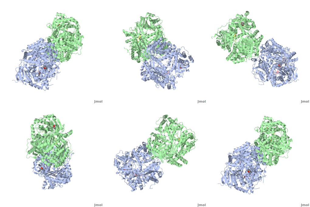
3D structures of insulin-degrading enzyme
Updated on 29-August-2019
References
- Im H, Manolopoulou M, Malito E, Shen Y, Zhao J, Neant-Fery M, Sun CY, Meredith SC, Sisodia SS, Leissring MA, Tang WJ. Structure of substrate-free human insulin-degrading enzyme (IDE) and biophysical analysis of ATP-induced conformational switch of IDE. J Biol Chem. 2007 Aug 31;282(35):25453-63. Epub 2007 Jul 5. PMID:17613531 doi:10.1074/jbc.M701590200
- Shen Y, Joachimiak A, Rosner MR, Tang WJ. Structures of human insulin-degrading enzyme reveal a new substrate recognition mechanism. Nature. 2006 Oct 19;443(7113):870-4. Epub 2006 Oct 11. PMID:17051221 doi:10.1038/nature05143
- Li P, Kuo WL, Yousef M, Rosner MR, Tang WJ. The C-terminal domain of human insulin degrading enzyme is required for dimerization and substrate recognition. Biochem Biophys Res Commun. 2006 May 19;343(4):1032-7. Epub 2006 Mar 22. PMID:16574064 doi:10.1016/j.bbrc.2006.03.083
- ↑ Chen KC, Chang SS, Tsai FJ, Chen CY. Han ethnicity-specific type 2 diabetic treatment from traditional Chinese medicine? J Biomol Struct Dyn. 2012 Nov 12. PMID:23146021 doi:10.1080/07391102.2012.732340
- ↑ Leissring MA, Malito E, Hedouin S, Reinstatler L, Sahara T, Abdul-Hay SO, Choudhry S, Maharvi GM, Fauq AH, Huzarska M, May PS, Choi S, Logan TP, Turk BE, Cantley LC, Manolopoulou M, Tang WJ, Stein RL, Cuny GD, Selkoe DJ. Designed inhibitors of insulin-degrading enzyme regulate the catabolism and activity of insulin. PLoS One. 2010 May 7;5(5):e10504. PMID:20498699 doi:10.1371/journal.pone.0010504
Proteopedia Page Contributors and Editors (what is this?)
Michal Harel, Alexander Berchansky, Élodie Weider, Karsten Theis, Joel L. Sussman, Jaime Prilusky

