Cystic fibrosis transmembrane conductance regulator (CFTR)
From Proteopedia
(Difference between revisions)
m |
|||
| Line 5: | Line 5: | ||
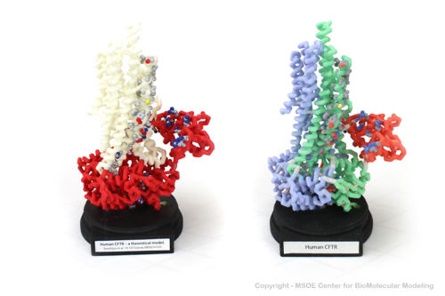
CFTR is a mostly <scene name='78/785332/Secondary_structure/1'>alpha helical</scene> protein. The membrane spanning segments can be clearly seen with coloring by <scene name='78/785332/Hydrophobicity/1'>hydrophobicity</scene>, which shows hydrophobic residues in gray and hydrophilic residues in purple. | CFTR is a mostly <scene name='78/785332/Secondary_structure/1'>alpha helical</scene> protein. The membrane spanning segments can be clearly seen with coloring by <scene name='78/785332/Hydrophobicity/1'>hydrophobicity</scene>, which shows hydrophobic residues in gray and hydrophilic residues in purple. | ||
| - | The extracellular end of the channel has several <scene name='78/785332/Ec_cl_selection/1'>positively charged</scene> residues that are important for recruiting chloride ions to the channel. A number of <scene name='78/785332/Plus_channel/1'>positively charged</scene> residues line the channel. In the unphosphorylated state (as this structure is), a <scene name='78/785332/Regulatory_domain/2'>regulatory domain</scene> blocks the activity of the channel (the connecting segments are not visible in the structure). It contains several negatively charged residues; when the protein is phosphorylated, this segment is repelled, causing a structural change. <ref>PMID: 28340353</ref> | + | The extracellular end of the channel has several <scene name='78/785332/Ec_cl_selection/1'>positively charged</scene> residues that are important for recruiting chloride ions to the channel. A number of <scene name='78/785332/Plus_channel/1'>positively charged</scene> residues line the channel. In the unphosphorylated state (as this structure is), a <scene name='78/785332/Regulatory_domain/2'>regulatory domain</scene> blocks the activity of the channel (the connecting segments are not visible in the structure). It contains several negatively charged residues; when the protein is phosphorylated, this segment is repelled, causing a structural change. <ref>PMID:28340353</ref> |
| - | + | ||
| + | CFTR contains two Walker motifs, which bind to nucleotides. | ||
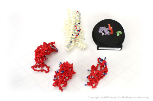
==3D Printed Physical Model of the CFTR protein== | ==3D Printed Physical Model of the CFTR protein== | ||
Revision as of 21:22, 16 October 2019
Cystic fibrosis transmembrane conductance regulator (CFTR)
| |||||||||||
References
- ↑ Liu F, Zhang Z, Csanady L, Gadsby DC, Chen J. Molecular Structure of the Human CFTR Ion Channel. Cell. 2017 Mar 23;169(1):85-95.e8. doi: 10.1016/j.cell.2017.02.024. PMID:28340353 doi:http://dx.doi.org/10.1016/j.cell.2017.02.024


