Cystic fibrosis transmembrane conductance regulator (CFTR)
From Proteopedia
| Line 8: | Line 8: | ||
CFTR contains two <scene name='78/785332/Nbd/2'>nucleotide binding domains</scene> (NBD's), which both contain <scene name='78/785332/Walker_motifs/1'>Walker motifs</scene>, flexible loops that bind phosphate groups tightly. | CFTR contains two <scene name='78/785332/Nbd/2'>nucleotide binding domains</scene> (NBD's), which both contain <scene name='78/785332/Walker_motifs/1'>Walker motifs</scene>, flexible loops that bind phosphate groups tightly. | ||
| + | |||
| + | ==Mutations in Cystic Fibrosis== | ||
| + | |||
| + | Cystic fibrosis is characterized by decreased chloride transport, which causes mucus to be thicker and stickier. This leads to a variety of problems, including decreased lung capacity, decreased pancreatic enzyme release into the small intestine, increased rates of lung infections, and infertility.<ref>https://ghr.nlm.nih.gov/condition/cystic-fibrosis<\ref> There are a wide assortment of mutations that cause cystic fibrosis, with differing symptom severity. The deletion of <scene name='78/785332/F508/1'>F508</scene> causes the protein to not be properly synthesized, and no expression is seen on the cell surface. | ||
==3D Printed Physical Model of the CFTR protein== | ==3D Printed Physical Model of the CFTR protein== | ||
Revision as of 21:53, 16 October 2019
Cystic fibrosis transmembrane conductance regulator (CFTR)
| |||||||||||
References
- ↑ Liu F, Zhang Z, Csanady L, Gadsby DC, Chen J. Molecular Structure of the Human CFTR Ion Channel. Cell. 2017 Mar 23;169(1):85-95.e8. doi: 10.1016/j.cell.2017.02.024. PMID:28340353 doi:http://dx.doi.org/10.1016/j.cell.2017.02.024
- ↑ https://ghr.nlm.nih.gov/condition/cystic-fibrosis<\ref> There are a wide assortment of mutations that cause cystic fibrosis, with differing symptom severity. The deletion of causes the protein to not be properly synthesized, and no expression is seen on the cell surface.
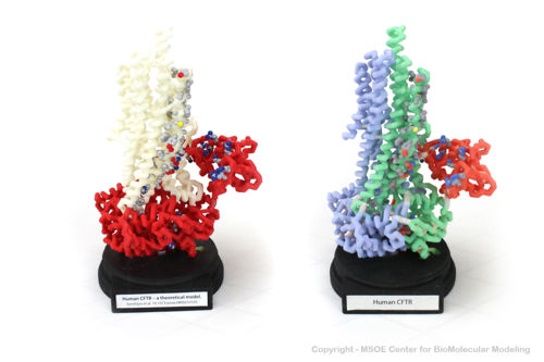
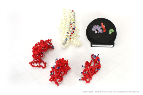
3D Printed Physical Model of the CFTR protein
Shown below are 3D printed physical models of the Cystic Fibrosis Transmembrane Conductance Regulator (CFTR) protein. The backbone model on the left is colored by region, with the transmembrane domain white and the atp-binding and regulatory domains colored red. The backbone model on the right is colored by regional repeat, with the first repeat blue, the second repeat green and the regulatory domain colored red. The models have been designed with embedded magnets to disassemble into the key regions of the structure.
The MSOE Center for BioMolecular Modeling

The MSOE Center for BioMolecular Modeling uses 3D printing technology to create physical models of protein and molecular structures, making the invisible molecular world more tangible and comprehensible. To view more protein structure models, visit our Model Gallery.


