DNA in action
From Proteopedia
m (Sandbox/Huntingtin moved to Sandbox over redirect) |
|||
| Line 1: | Line 1: | ||
| - | #REDIRECT [[ | + | == IPEX syndrome == |
| + | |||
| + | ---- | ||
| + | Immune dysregulation, polyendocrinopathy, entheropathy, X-linked syndrome is rare autoimmune disorder caused by genetic mutation in FOXP3 gene, which is responsible for producing important transcription factor required for maintenance of T regulatory cells (T-regs). T-reg cells disfunction is main pathogenic event which leads to multiorgan autoimmunity called IPEX syndrome. <ref>Bacchetta, R., Barzaghi, F., & Roncarolo, M.-G. (2016). From IPEX syndrome to FOXP3 mutation: a lesson on immune dysregulation. Annals of the New York Academy of Sciences, 1417(1), 5–22. doi:10.1111/nyas.13011 </ref> | ||
| + | |||
| + | '''FOXP3 gene structure''' | ||
| + | ---- | ||
| + | FOXP3 gene is located on centromeric region of the X chromosome in position Xq11.3-q13.3 and is comprised of 12 exones. Molecular location is from 49,250,436- 49,264,932 base pairs on the X chromosome. This gene can be also found under other names such as: AIID, DIETER, IPEX, scurfin… In humans is this highly conserved gene responsible for production of 431 amino acids long fork head protein P3 (FOXP3 protein). This protein is a member of the FKH family of transcription factors. Contains proline rich (PRR) aminoterminal domain, central zinc finger (ZC), leucine zipper domain and carboxyl terminal FKH domain. | ||
| + | |||
| + | '''Function''' | ||
| + | ---- | ||
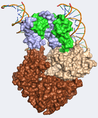
| + | Probably the most characteristic part of FOXP3 protein is the “forkhead box”. This part of the protein, consisting of 80-100 amino acids, form a DNA binding motif, giving FOXP3 protein capacity to bind DNA and regulate expression of various genes and is highly expressed in CD4+CD25+ regulatory T cells (T-regs). FOXP3 is able bind to a number of distinct genomic loci in T-regs and therefore can function as both repressor and trans-activator depending on the interaction partners. This specific T cell subset is involved in limiting the immune response of other cells, for example conventional cytotoxic T cells. T-regs are developed from relatively small population of CD4+T helper cells in thymus. They are characterized by surface markers such as CD25 or CTLA4, but most importantly FOXP3 which is functionally involved in immunosuppression and therefore without FOXP3 protein present T-reg cells do not develop at all. | ||
| + | |||
| + | |||
| + | [[Image:Picturefoxp34.png]] | ||
| + | |||
| + | For more information on structure and function see also #REDIRECT [[Forkhead Box Protein 3]] | ||
| + | |||
| + | '''Mutations''' | ||
| + | ---- | ||
| + | For development of IPEX syndrome at least 70 distinct FOXP3 mutations have been reported. However, among deferent patient same mutation can be responsible for different phenotypes. For example, mutation in FKH domain (c.1150G>A) is connected with patients surviving more than 10 years, but there are also reported cases when patient died prematurely. Out of all identified mutations 40% reside in C-terminal FKH DNA binding domain, 23% in N-terminal PRR domain, 9% in the LZ domain, 16% in the LZ-FKH loop, 6% in the noncoding region upstream of initiating ATG and last 6% in C- terminal end of the ORF. Most commons among patients are missense mutations, mostly point mutations, that are responsible for normal or reduced expression of mutant protein. Severe phenotype was usually observed at individuals with completely impaired expression of functional FOXP3 protein (frameshift, missplicing…). | ||
| + | |||
| + | '''IPEX phenotype and clinical manifestations''' | ||
| + | ---- | ||
| + | In most cases IPEX are born at term with normal weight and length. However, first signs of illness may present itself immediately after birth or in the very first days of life. This suggests that autoimmune process has been already initiated in utero. Some case reportedly developed intrauterine growth retardation and shortly after birth they developed also standard IPEX phenotype. Severe symptoms with early onset can be rapidly fatal with absence of early diagnostics and treatment. Most common symptoms are diarrhea, type 1 diabetes and eczema, however overall picture can be complicated with other autoimmune symptoms such as. Development of Autoimmune enteropathy is one of the key hallmarks of the disease presenting itself as neonatal watery diarrhea with traces of mucus and blood. This symptom starts with breastfeeding and usually worsens when baby starts implementing regular nutrition which may result in severe malabsorption. Type one diabetes can precede or follow enteritis. Similar to diabetes and diarrhea cutaneous manifestations are also very common and may present shortly after birth and can range from mild dermatitis to severe and diffuse lesions. These lesions might be accompanied by bacterial infections. Other symptoms that may be present are thyroid dysfunction, autoimmune cytopenia, autoimmune hemolytic anemia, renal disease, arthritis (involving one or more joints). | ||
| + | |||
| + | '''Diagnostics''' | ||
| + | ---- | ||
| + | For a correct diagnosis family history is of a great value. However, the symptoms of the disease may vary within family members. This may be caused by different regulatory elements, differences in gene modifications, different environment or previous treatment. Among clinical symptoms signs of type one diabetes, hypothyroidism, enteropathy and cytopenia may be present. Histopathology analysis shows complete absence of normal mucosa in the small bowel and colon sometimes with infiltrations of inflammatory cells in the lamina propria and submucosa as well as in other organs including pancreas, skin or kidneys. From the blood autoantibodies can be detected against pancreas, thyroid and erythrocytes. Changes in complement elements, granulocytes conventional T cells or immunoglobulins have not been reported. This however may not be enough for disease confirmation. For final differential diagnostics has to be based on DNA analysis which confirms mutations in FOXP3 gene. | ||
| + | |||
| + | '''Treatment''' | ||
| + | ---- | ||
| + | As for now, the most promising treatment comes from combination of immunosuppressive therapy with bone marrow transplantation with addition of supportive treatment including parenteral nutrition. IPEX syndrome is also a good candidate for gene therapy treatment, however, still needs more preclinical studies to evaluate safety and efficiency. | ||
| + | |||
| + | '''References''' | ||
| + | ---- | ||
| + | <references/> | ||
Revision as of 20:28, 30 April 2020
IPEX syndrome
Immune dysregulation, polyendocrinopathy, entheropathy, X-linked syndrome is rare autoimmune disorder caused by genetic mutation in FOXP3 gene, which is responsible for producing important transcription factor required for maintenance of T regulatory cells (T-regs). T-reg cells disfunction is main pathogenic event which leads to multiorgan autoimmunity called IPEX syndrome. [1]
FOXP3 gene structure
FOXP3 gene is located on centromeric region of the X chromosome in position Xq11.3-q13.3 and is comprised of 12 exones. Molecular location is from 49,250,436- 49,264,932 base pairs on the X chromosome. This gene can be also found under other names such as: AIID, DIETER, IPEX, scurfin… In humans is this highly conserved gene responsible for production of 431 amino acids long fork head protein P3 (FOXP3 protein). This protein is a member of the FKH family of transcription factors. Contains proline rich (PRR) aminoterminal domain, central zinc finger (ZC), leucine zipper domain and carboxyl terminal FKH domain.
Function
Probably the most characteristic part of FOXP3 protein is the “forkhead box”. This part of the protein, consisting of 80-100 amino acids, form a DNA binding motif, giving FOXP3 protein capacity to bind DNA and regulate expression of various genes and is highly expressed in CD4+CD25+ regulatory T cells (T-regs). FOXP3 is able bind to a number of distinct genomic loci in T-regs and therefore can function as both repressor and trans-activator depending on the interaction partners. This specific T cell subset is involved in limiting the immune response of other cells, for example conventional cytotoxic T cells. T-regs are developed from relatively small population of CD4+T helper cells in thymus. They are characterized by surface markers such as CD25 or CTLA4, but most importantly FOXP3 which is functionally involved in immunosuppression and therefore without FOXP3 protein present T-reg cells do not develop at all.
For more information on structure and function see also #REDIRECT Forkhead Box Protein 3
Mutations
For development of IPEX syndrome at least 70 distinct FOXP3 mutations have been reported. However, among deferent patient same mutation can be responsible for different phenotypes. For example, mutation in FKH domain (c.1150G>A) is connected with patients surviving more than 10 years, but there are also reported cases when patient died prematurely. Out of all identified mutations 40% reside in C-terminal FKH DNA binding domain, 23% in N-terminal PRR domain, 9% in the LZ domain, 16% in the LZ-FKH loop, 6% in the noncoding region upstream of initiating ATG and last 6% in C- terminal end of the ORF. Most commons among patients are missense mutations, mostly point mutations, that are responsible for normal or reduced expression of mutant protein. Severe phenotype was usually observed at individuals with completely impaired expression of functional FOXP3 protein (frameshift, missplicing…).
IPEX phenotype and clinical manifestations
In most cases IPEX are born at term with normal weight and length. However, first signs of illness may present itself immediately after birth or in the very first days of life. This suggests that autoimmune process has been already initiated in utero. Some case reportedly developed intrauterine growth retardation and shortly after birth they developed also standard IPEX phenotype. Severe symptoms with early onset can be rapidly fatal with absence of early diagnostics and treatment. Most common symptoms are diarrhea, type 1 diabetes and eczema, however overall picture can be complicated with other autoimmune symptoms such as. Development of Autoimmune enteropathy is one of the key hallmarks of the disease presenting itself as neonatal watery diarrhea with traces of mucus and blood. This symptom starts with breastfeeding and usually worsens when baby starts implementing regular nutrition which may result in severe malabsorption. Type one diabetes can precede or follow enteritis. Similar to diabetes and diarrhea cutaneous manifestations are also very common and may present shortly after birth and can range from mild dermatitis to severe and diffuse lesions. These lesions might be accompanied by bacterial infections. Other symptoms that may be present are thyroid dysfunction, autoimmune cytopenia, autoimmune hemolytic anemia, renal disease, arthritis (involving one or more joints).
Diagnostics
For a correct diagnosis family history is of a great value. However, the symptoms of the disease may vary within family members. This may be caused by different regulatory elements, differences in gene modifications, different environment or previous treatment. Among clinical symptoms signs of type one diabetes, hypothyroidism, enteropathy and cytopenia may be present. Histopathology analysis shows complete absence of normal mucosa in the small bowel and colon sometimes with infiltrations of inflammatory cells in the lamina propria and submucosa as well as in other organs including pancreas, skin or kidneys. From the blood autoantibodies can be detected against pancreas, thyroid and erythrocytes. Changes in complement elements, granulocytes conventional T cells or immunoglobulins have not been reported. This however may not be enough for disease confirmation. For final differential diagnostics has to be based on DNA analysis which confirms mutations in FOXP3 gene.
Treatment
As for now, the most promising treatment comes from combination of immunosuppressive therapy with bone marrow transplantation with addition of supportive treatment including parenteral nutrition. IPEX syndrome is also a good candidate for gene therapy treatment, however, still needs more preclinical studies to evaluate safety and efficiency.
References
- ↑ Bacchetta, R., Barzaghi, F., & Roncarolo, M.-G. (2016). From IPEX syndrome to FOXP3 mutation: a lesson on immune dysregulation. Annals of the New York Academy of Sciences, 1417(1), 5–22. doi:10.1111/nyas.13011
Proteopedia Page Contributors and Editors (what is this?)
Emily Joyce, Brian Boyle, Hannah Campbell, Antoine Lambert, Elizeu Santos, Ivan Šonský, Adrián Šutta, Atena Farhangian, Vishal Bhoir

