This old version of Proteopedia is provided for student assignments while the new version is undergoing repairs. Content and edits done in this old version of Proteopedia after March 1, 2026 will eventually be lost when it is retired in about June of 2026.
Apply for new accounts at the new Proteopedia. Your logins will work in both the old and new versions.
Galactose-binding lectin
From Proteopedia
(Difference between revisions)
| Line 76: | Line 76: | ||
**[[2ds0]] – LtGBL C terminal (mutant) + sialyllactose <br /> | **[[2ds0]] – LtGBL C terminal (mutant) + sialyllactose <br /> | ||
**[[2zqo]] – LtGBL C terminal + GalNac <br /> | **[[2zqo]] – LtGBL C terminal + GalNac <br /> | ||
| + | |||
| + | *Other GBL | ||
| + | |||
| + | **[[5x4a]], [[3wmp]], [[3wmq]] – GBL residues 47-140 + polysaccharide – ''Sinularia lochmodes'' <br /> | ||
| + | **[[2zqn]] – eGBL C-terminal + lactose – earthworm<br /> | ||
| + | **[[2zqo]] – eGBL C-terminal + GalNac<br /> | ||
}} | }} | ||
== References == | == References == | ||
<references/> | <references/> | ||
[[Category:Topic Page]] | [[Category:Topic Page]] | ||
Revision as of 09:38, 30 July 2020
| |||||||||||
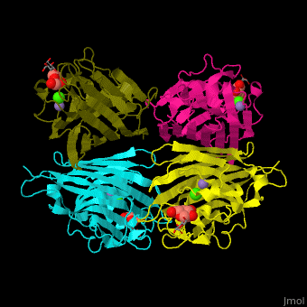
3D structures of galactose-binding lectin
Updated on 30-July-2020
References
- ↑ Natchiar SK, Srinivas O, Mitra N, Surolia A, Jayaraman N, Vijayan M. Structural studies on peanut lectin complexed with disaccharides involving different linkages: further insights into the structure and interactions of the lectin. Acta Crystallogr D Biol Crystallogr. 2006 Nov;62(Pt 11):1413-21. Epub 2006, Oct 18. PMID:17057347 doi:10.1107/S0907444906035712
- ↑ Kundhavai Natchiar S, Arockia Jeyaprakash A, Ramya TN, Thomas CJ, Suguna K, Surolia A, Vijayan M. Structural plasticity of peanut lectin: an X-ray analysis involving variation in pH, ligand binding and crystal structure. Acta Crystallogr D Biol Crystallogr. 2004 Feb;60(Pt 2):211-9. Epub 2004, Jan 23. PMID:14747696 doi:http://dx.doi.org/10.1107/S090744490302849X
