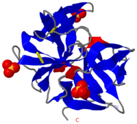
Chymotrypsin
From Proteopedia
| Line 19: | Line 19: | ||
This view shows the <scene name='38/387136/Bovine_chymotrypsin_active_sit/3'>carbonyl group of the inhibitor</scene> in CPK colors. The triflouromethyl group is bound to the carbonyl carbon via the yellow bond. In a peptide substrate, the triflouromethyl group would be replaced by the first amino acid residue of the rest of the peptide chain, and the yellow bond would be the bond that is cleaved. The carbonyl carbon of the inhibitor is 1.52 Å away from the side chain oxygen of serine 195, and this indicates they are covalently bound (bond indicated by dotted line). Thus, this structure is similar to the '''tetrahedral intermediate''' that is formed during the cleavage reaction. The negative charge that develops on the carbonyl oxygen of the substrate is stabilized by hydrogen bonds to the backbone nitrogens of Ser 195 and Gly 193, shown in blue spacefill. The hydrogen atoms involved in these hydrogen bonds are not shown. | This view shows the <scene name='38/387136/Bovine_chymotrypsin_active_sit/3'>carbonyl group of the inhibitor</scene> in CPK colors. The triflouromethyl group is bound to the carbonyl carbon via the yellow bond. In a peptide substrate, the triflouromethyl group would be replaced by the first amino acid residue of the rest of the peptide chain, and the yellow bond would be the bond that is cleaved. The carbonyl carbon of the inhibitor is 1.52 Å away from the side chain oxygen of serine 195, and this indicates they are covalently bound (bond indicated by dotted line). Thus, this structure is similar to the '''tetrahedral intermediate''' that is formed during the cleavage reaction. The negative charge that develops on the carbonyl oxygen of the substrate is stabilized by hydrogen bonds to the backbone nitrogens of Ser 195 and Gly 193, shown in blue spacefill. The hydrogen atoms involved in these hydrogen bonds are not shown. | ||
| + | |||
| + | |||
| + | </StructureSection> | ||
| + | |||
| + | == Mechanism == | ||
| + | The hydrolysis occurs in two steps. First, the peptide bond with the C-terminal part of the substrate is replaced by an ester bond with the active site serin. Second, the covalent intermediate is hydrolyzed by water, releasing the N-terminal part of the substrate as carboxylic acid. Both steps occur via a tetrahedral intermediate containing an oxyanion that is stabilized by hydrogen bond donors lining the so-called oxyanion hole. Throughout the reaction, active site residue histidine 57 acts as base or acid to deprotonate nucleophiles or protonate leaving groups, respectively. The following 5-minute video shows the mechanism step by step. | ||
| + | |||
| + | <center><html5media height=“315” width=“560” frameborder="0" allowfullscreen>https://www.youtube.com/embed/X3HmYhR2BA0</html5media></center> | ||
| + | <center>Mechanism of chymotrypsin </center> | ||
| + | |||
== 3D Structures of Chymotrypsin == | == 3D Structures of Chymotrypsin == | ||
[[Chymotrypsin 3D structures]] | [[Chymotrypsin 3D structures]] | ||
| - | </StructureSection> | ||
== References == | == References == | ||
Revision as of 21:54, 11 October 2020
| |||||||||||
Contents |
Mechanism
The hydrolysis occurs in two steps. First, the peptide bond with the C-terminal part of the substrate is replaced by an ester bond with the active site serin. Second, the covalent intermediate is hydrolyzed by water, releasing the N-terminal part of the substrate as carboxylic acid. Both steps occur via a tetrahedral intermediate containing an oxyanion that is stabilized by hydrogen bond donors lining the so-called oxyanion hole. Throughout the reaction, active site residue histidine 57 acts as base or acid to deprotonate nucleophiles or protonate leaving groups, respectively. The following 5-minute video shows the mechanism step by step.
3D Structures of Chymotrypsin
References
- ↑ Appel W. Chymotrypsin: molecular and catalytic properties. Clin Biochem. 1986 Dec;19(6):317-22. PMID:3555886
Further reading
You can learn more about chymotrypsin structure, function and regulation in this publicly available chapter of the Biochemistry textbook by Berg, Tymoczka and Stryer.
Proteopedia Page Contributors and Editors (what is this?)
Michal Harel, Karsten Theis, Alice Harmon, Alexander Berchansky


