Introduction
G protein-coupled receptors (GPCRs) are a large family of protein receptors, which have seven-transmembrane helices and are found over a large array of eukaryotic cells. These receptors take a major part in a multitude of signal transduction pathways, including amongst others responses to hormones and neurotransmitters, sensing light, taste and smell, and many more. These receptors are also involved in many different types of diseases and are the target of almost 50% of current medical drugs.
The Beta-2 Adrenergic Receptors are a type of GPCR that are activated by catecholamine hormone ligands such as adrenaline (epinephrine). These receptors are responsible for many of the adrenaline related (“fight-or-flight”) responses and functions, and are used as a common model system for the GPCR family.
GPCRs bind their ligand and overcome a conformational change that activates an attached Guanine nucleotide-binding protein (G protein) and allows it to detach from the cellular end of the receptor and start the different signal transduction pathways.
G proteins[1] are a family of proteins that act as molecular switches inside cells. G proteins belong to the larger group of enzymes called GTPases[2], and appear either as monomeric small GTPases[3], or as heterotrimeric G protein complexes[4] that are made up of alpha (α), beta (β) and gamma (γ) subunits[5]. When they are bound to guanosine triphosphate (GTP), they are 'on', and when they are bound to guanosine diphosphate (GDP), they are 'off'.
Since these receptors have seven transmembrane helices as well as inner and outer cell regions, they are very difficult to purify and crystallize. Some crystal structures have been determined for the inactive receptors as well as for the G proteins that they bind. PDB entry 3SN6 is the first structure of the full complex of the Beta 2 Adrenergic Receptor bound to Gs in their active state, and it provides the first high-resolution insight into the mechanism of signal transduction across the plasma membrane by a GPCR.
Complex structure
The overall structure shows the (dark blue) bound to an agonist (in spheres) along with a lysozyme fused to its amino terminus in order to facilitate crystallization. The receptor interacts with (light blue). Gαs together with (light green) and (gold) constitute the heterotrimeric G protein Gs. A Gs-binding nanobody , which also facilitates crystallization, binds the G protein between the α and β subunits.
G-Protein-GPCR Interactions
The α5-helix of Gαs into a cavity formed on the intracellular side of the receptor by the opening of the transmembrane helices. Within the transmembrane core, the interactions are primarily non-polar - an exception involves of Tyr 391 of the α5-helix against Arg 131 of the conserved DRY sequence in TM3. Arg 131 also packs against Tyr 326 of the conserved NPxxY sequence in TM7. As the α5-helix exits the receptor it forms a network of polar interactions with TM5 and TM3. Receptor residues Thr 68 and Asp 130 interact with the ICL2 helix of the β2AR via Tyr 141, positioning the helix so that Phe 139 of the receptor a hydrophobic pocket on the G protein surface, thereby structurally linking receptor–G protein interactions with the highly conserved DRY motif of the β2AR.
G-Protein Activation Cycle
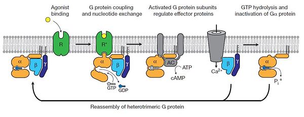
The figure shows the G Protein cycle[6] - an extracellular agonist binding to the β2AR leads to of the cytoplasmic ends of transmembrane segments that enable the Gs heterotrimer to bind the receptor. GDP is released from the α subunit upon formation of β2AR–Gs complex. The GTP binds to the nucleotide-free α subunit resulting in dissociation of the α and βγ subunits from the receptor. The subunits regulate their respective effector proteins adenylyl cyclase (AC) and Ca2+ channels. The Gs heterotrimer reassembles from α and βγ subunits following hydrolysis of GTP to GDP in the α subunit.