We apologize for Proteopedia being slow to respond. For the past two years, a new implementation of Proteopedia has been being built. Soon, it will replace this 18-year old system. All existing content will be moved to the new system at a date that will be announced here.
User:Megan Leaman/Sandbox 1
From Proteopedia
(Difference between revisions)
| Line 20: | Line 20: | ||
=== Mechanism === | === Mechanism === | ||
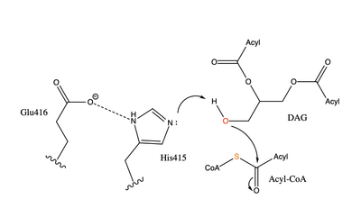
| - | [[Image:dgat chemdraw mechanism.png|400 px|right|thumb|Figure 4]]The catalytic deprotonates the hydroxyl group on the C3 of the glycerol backbone. The deprotonated oxygen then makes a nucleophilic attack on the carbonyl carbon of the Acyl-CoA, the electron density gets shifted up to the oxygen and back down to the carbonyl carbon. The sulfur of the Acyl-CoA takes the added electron density and the bond between the sulfur and carbonyl carbon is broken. Glu416 likely provides transition state stabilization of the His415 <ref name="Sui"></ref> despite being outside of the 3 angstrom hydrogen binding distance. This is likely due to the fact that the DGAT 1 enzyme was unable to be visualized with the diacylglycerol in the active site. The entrance and binding of the diacylglycerol may cause conformational changes and shifting of the Glu416 to become closer to the His415. Point mutations made to His415 support the hypothesis that it is essential for stabilization in the active site since the enzyme function was completely eliminated when the mutation was made. | + | [[Image:dgat chemdraw mechanism.png|400 px|right|thumb|Figure 4]]The catalytic deprotonates the hydroxyl group on the C3 of the glycerol backbone. The deprotonated oxygen then makes a nucleophilic attack on the carbonyl carbon of the Acyl-CoA, the electron density gets shifted up to the oxygen and back down to the carbonyl carbon. The sulfur of the Acyl-CoA takes the added electron density and the bond between the sulfur and carbonyl carbon is broken. Glu416 likely provides transition state stabilization of the His415 <ref name="Sui"> Sui, X., Wang, K., Gluchowski, N. L., Elliott, S. D., Liao, M., Walther, T. C., & Farese, R. V. (2020). Structure and catalytic mechanism of a human triacylglycerol-synthesis enzyme. Nature, 581(7808), 323-328. doi:10.1038/s41586-020-2289-6</ref> despite being outside of the 3 angstrom hydrogen binding distance. This is likely due to the fact that the DGAT 1 enzyme was unable to be visualized with the diacylglycerol in the active site. The entrance and binding of the diacylglycerol may cause conformational changes and shifting of the Glu416 to become closer to the His415. Point mutations made to His415 support the hypothesis that it is essential for stabilization in the active site since the enzyme function was completely eliminated when the mutation was made. |
While there is not universal agreement on whether <scene name='87/878228/Gln465_vs_glu416/3'>Gln465 or Glu416</scene> provide transition state stabilization for the acyl transfer activity of the enzyme, it is believed that Glu416 is more likely to provide stabilization due to positive findings upon mutations of that residue. Point mutations were made to various residues in the active site including Glu416. Mutations to the entrance of the binding site have a smaller impact on the functionality of the enzyme than mutation to the rest of the active site, while mutations to His415 and Glu416 abolish enzymatic activity completely. This suggests that His415 and Glu416 are essential to the reaction whereas Gln465 can be changed without significantly impacting the functionality of the enzyme. | While there is not universal agreement on whether <scene name='87/878228/Gln465_vs_glu416/3'>Gln465 or Glu416</scene> provide transition state stabilization for the acyl transfer activity of the enzyme, it is believed that Glu416 is more likely to provide stabilization due to positive findings upon mutations of that residue. Point mutations were made to various residues in the active site including Glu416. Mutations to the entrance of the binding site have a smaller impact on the functionality of the enzyme than mutation to the rest of the active site, while mutations to His415 and Glu416 abolish enzymatic activity completely. This suggests that His415 and Glu416 are essential to the reaction whereas Gln465 can be changed without significantly impacting the functionality of the enzyme. | ||
Revision as of 13:03, 20 April 2021
Human Diacylglycerol O-Transferase 1
| |||||||||||
References
- ↑ 1.0 1.1 Cases S, Smith SJ, Zheng YW, Myers HM, Lear SR, Sande E, Novak S, Collins C, Welch CB, Lusis AJ, Erickson SK, Farese RV Jr. Identification of a gene encoding an acyl CoA:diacylglycerol acyltransferase, a key enzyme in triacylglycerol synthesis. Proc Natl Acad Sci U S A. 1998 Oct 27;95(22):13018-23. PMID:9789033
- ↑ 2.0 2.1 Yen CL, Stone SJ, Koliwad S, Harris C, Farese RV Jr. Thematic review series: glycerolipids. DGAT enzymes and triacylglycerol biosynthesis. J Lipid Res. 2008 Nov;49(11):2283-301. doi: 10.1194/jlr.R800018-JLR200. Epub 2008, Aug 29. PMID:18757836 doi:http://dx.doi.org/10.1194/jlr.R800018-JLR200
- ↑ 3.0 3.1 Sui, X., Wang, K., Gluchowski, N. L., Elliott, S. D., Liao, M., Walther, T. C., & Farese, R. V. (2020). Structure and catalytic mechanism of a human triacylglycerol-synthesis enzyme. Nature, 581(7808), 323-328. doi:10.1038/s41586-020-2289-6
- ↑ Ransey E, Paredes E, Dey SK, Das SR, Heroux A, Macbeth MR. Crystal structure of the Entamoeba histolytica RNA lariat debranching enzyme EhDbr1 reveals a catalytic Zn(2+) /Mn(2+) heterobinucleation. FEBS Lett. 2017 Jul;591(13):2003-2010. doi: 10.1002/1873-3468.12677. Epub 2017, Jun 14. PMID:28504306 doi:http://dx.doi.org/10.1002/1873-3468.12677
- ↑ Wang L;Qian H;Nian Y;Han Y;Ren Z;Zhang H;Hu L;Prasad BVV;Laganowsky A;Yan N;Zhou M;. (2020, May 13). Structure and mechanism of human diacylglycerol o-acyltransferase 1. Retrieved March 09, 2021, from https://pubmed.ncbi.nlm.nih.gov/32433610/
Student Contributors
- Megan Leaman
- Grace Hall
- Karina Latsko


