User:Brianna Avery/Sandbox 1
From Proteopedia
(Difference between revisions)
| Line 20: | Line 20: | ||
====Metal Cations==== | ====Metal Cations==== | ||
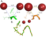
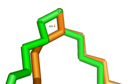
| - | [[Image: | + | [[Image:Best_Metal_Cation_Figure.png|150 px|right|thumb|'''Differences between zinc and iron di-metal interactions with acetyl-CoA.''']]Typically, SCD1 contains a <scene name='87/877504/Di_metal_center/2'>di-metal center</scene> in the core of the protein that contributes to its desaturation function. Crystallized structures of SCD1 indicated a substitution of Zinc as the di-metal center instead of <ref name="Bai" />. It is proposed that the difference in size of Zinc and Iron (8.8 Å and 9.2 Å respectively) would not affect the structure of SCD (Figure containing all 3 of Treys photos?). However, the substitution of Zinc as the di-iron center is found to inhibit the function of SCD1 due to a farther distance between the two metals compared to iron (Figure containing all 3 of Treys photos) <ref name="Bai" />. |
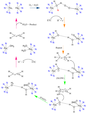
In other di-metal centered enzymes such as [https://en.wikipedia.org/wiki/Acyl-(acyl-carrier-protein)_desaturase ACP desaturase] and [https://en.wikipedia.org/wiki/Ribonucleotide_reductase ribonucleotide reductase], the enzymatic mechanism involves an oxo-bridge: a water molecule that is recruited by the di-iron center to be directly involved in the desaturase mechanism. This water gets deprotonated by the two metal ions and it becomes nucleophilic enough to attack the substrate (Mechanism figure). Based on electron density mapping of SCD1, this oxo-bridge formation is suggested to be short-lived <ref name="Shen">DOI: 10.1016/j.jmb.2020.05.017</ref>. | In other di-metal centered enzymes such as [https://en.wikipedia.org/wiki/Acyl-(acyl-carrier-protein)_desaturase ACP desaturase] and [https://en.wikipedia.org/wiki/Ribonucleotide_reductase ribonucleotide reductase], the enzymatic mechanism involves an oxo-bridge: a water molecule that is recruited by the di-iron center to be directly involved in the desaturase mechanism. This water gets deprotonated by the two metal ions and it becomes nucleophilic enough to attack the substrate (Mechanism figure). Based on electron density mapping of SCD1, this oxo-bridge formation is suggested to be short-lived <ref name="Shen">DOI: 10.1016/j.jmb.2020.05.017</ref>. | ||
Revision as of 15:32, 26 April 2021
Desaturation of Fatty Stearoyl-CoA by SCD1
| |||||||||||
References
- ↑ 1.0 1.1 1.2 1.3 1.4 1.5 1.6 1.7 Bai Y, McCoy JG, Levin EJ, Sobrado P, Rajashankar KR, Fox BG, Zhou M. X-ray structure of a mammalian stearoyl-CoA desaturase. Nature. 2015 Jun 22. doi: 10.1038/nature14549. PMID:26098370 doi:http://dx.doi.org/10.1038/nature14549
- ↑ 2.0 2.1 2.2 2.3 2.4 2.5 Tracz-Gaszewska Z, Dobrzyn P. Stearoyl-CoA Desaturase 1 as a Therapeutic Target for the Treatment of Cancer. Cancers (Basel). 2019 Jul 5;11(7). pii: cancers11070948. doi:, 10.3390/cancers11070948. PMID:31284458 doi:http://dx.doi.org/10.3390/cancers11070948
- ↑ 3.0 3.1 3.2 Shen J, Wu G, Tsai AL, Zhou M. Structure and Mechanism of a Unique Diiron Center in Mammalian Stearoyl-CoA Desaturase. J Mol Biol. 2020 May 27. pii: S0022-2836(20)30367-3. doi:, 10.1016/j.jmb.2020.05.017. PMID:32470559 doi:http://dx.doi.org/10.1016/j.jmb.2020.05.017
- ↑ Wang H, Klein MG, Zou H, Lane W, Snell G, Levin I, Li K, Sang BC. Crystal structure of human stearoyl-coenzyme A desaturase in complex with substrate. Nat Struct Mol Biol. 2015 Jul;22(7):581-5. doi: 10.1038/nsmb.3049. Epub 2015 Jun , 22. PMID:26098317 doi:http://dx.doi.org/10.1038/nsmb.3049
- ↑ 5.0 5.1 Gutierrez-Juarez R, Pocai A, Mulas C, Ono H, Bhanot S, Monia BP, Rossetti L. Critical role of stearoyl-CoA desaturase-1 (SCD1) in the onset of diet-induced hepatic insulin resistance. J Clin Invest. 2006 Jun;116(6):1686-95. doi: 10.1172/JCI26991. PMID:16741579 doi:http://dx.doi.org/10.1172/JCI26991
- ↑ Yokoyama S, Hosoi T, Ozawa K. Stearoyl-CoA Desaturase 1 (SCD1) is a key factor mediating diabetes in MyD88-deficient mice. Gene. 2012 Apr 15;497(2):340-3. doi: 10.1016/j.gene.2012.01.024. Epub 2012 Feb 3. PMID:22326531 doi:http://dx.doi.org/10.1016/j.gene.2012.01.024
- ↑ Ntambi JM, Miyazaki M, Stoehr JP, Lan H, Kendziorski CM, Yandell BS, Song Y, Cohen P, Friedman JM, Attie AD. Loss of stearoyl-CoA desaturase-1 function protects mice against adiposity. Proc Natl Acad Sci U S A. 2002 Aug 20;99(17):11482-6. doi:, 10.1073/pnas.132384699. Epub 2002 Aug 12. PMID:12177411 doi:http://dx.doi.org/10.1073/pnas.132384699
- ↑ Holder AM, Gonzalez-Angulo AM, Chen H, Akcakanat A, Do KA, Fraser Symmans W, Pusztai L, Hortobagyi GN, Mills GB, Meric-Bernstam F. High stearoyl-CoA desaturase 1 expression is associated with shorter survival in breast cancer patients. Breast Cancer Res Treat. 2013 Jan;137(1):319-27. doi: 10.1007/s10549-012-2354-4. , Epub 2012 Dec 4. PMID:23208590 doi:http://dx.doi.org/10.1007/s10549-012-2354-4
- ↑ Li J, Condello S, Thomes-Pepin J, Ma X, Xia Y, Hurley TD, Matei D, Cheng JX. Lipid Desaturation Is a Metabolic Marker and Therapeutic Target of Ovarian Cancer Stem Cells. Cell Stem Cell. 2017 Mar 2;20(3):303-314.e5. doi: 10.1016/j.stem.2016.11.004., Epub 2016 Dec 29. PMID:28041894 doi:http://dx.doi.org/10.1016/j.stem.2016.11.004
Student Contributors
- Brianna M. Avery
- William J. Harris III
- Emily M. Royston


