This old version of Proteopedia is provided for student assignments while the new version is undergoing repairs. Content and edits done in this old version of Proteopedia after March 1, 2026 will eventually be lost when it is retired in about June of 2026.
Apply for new accounts at the new Proteopedia. Your logins will work in both the old and new versions.
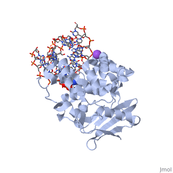
1diz
From Proteopedia
(Difference between revisions)
| Line 3: | Line 3: | ||
<StructureSection load='1diz' size='340' side='right'caption='[[1diz]], [[Resolution|resolution]] 2.50Å' scene=''> | <StructureSection load='1diz' size='340' side='right'caption='[[1diz]], [[Resolution|resolution]] 2.50Å' scene=''> | ||
== Structural highlights == | == Structural highlights == | ||
| - | <table><tr><td colspan='2'>[[1diz]] is a 6 chain structure with sequence from [ | + | <table><tr><td colspan='2'>[[1diz]] is a 6 chain structure with sequence from [https://en.wikipedia.org/wiki/"bacillus_coli"_migula_1895 "bacillus coli" migula 1895]. Full crystallographic information is available from [http://oca.weizmann.ac.il/oca-bin/ocashort?id=1DIZ OCA]. For a <b>guided tour on the structure components</b> use [https://proteopedia.org/fgij/fg.htm?mol=1DIZ FirstGlance]. <br> |
| - | </td></tr><tr id='ligand'><td class="sblockLbl"><b>[[Ligand|Ligands:]]</b></td><td class="sblockDat"><scene name='pdbligand=NA:SODIUM+ION'>NA</scene></td></tr> | + | </td></tr><tr id='ligand'><td class="sblockLbl"><b>[[Ligand|Ligands:]]</b></td><td class="sblockDat" id="ligandDat"><scene name='pdbligand=NA:SODIUM+ION'>NA</scene></td></tr> |
<tr id='NonStdRes'><td class="sblockLbl"><b>[[Non-Standard_Residue|NonStd Res:]]</b></td><td class="sblockDat"><scene name='pdbligand=NRI:PHOSPHORIC+ACID+MONO-(4-HYDROXY-PYRROLIDIN-3-YLMETHYL)+ESTER'>NRI</scene></td></tr> | <tr id='NonStdRes'><td class="sblockLbl"><b>[[Non-Standard_Residue|NonStd Res:]]</b></td><td class="sblockDat"><scene name='pdbligand=NRI:PHOSPHORIC+ACID+MONO-(4-HYDROXY-PYRROLIDIN-3-YLMETHYL)+ESTER'>NRI</scene></td></tr> | ||
| - | <tr id='activity'><td class="sblockLbl"><b>Activity:</b></td><td class="sblockDat"><span class='plainlinks'>[ | + | <tr id='activity'><td class="sblockLbl"><b>Activity:</b></td><td class="sblockDat"><span class='plainlinks'>[https://en.wikipedia.org/wiki/DNA-3-methyladenine_glycosylase_II DNA-3-methyladenine glycosylase II], with EC number [https://www.brenda-enzymes.info/php/result_flat.php4?ecno=3.2.2.21 3.2.2.21] </span></td></tr> |
| - | <tr id='resources'><td class="sblockLbl"><b>Resources:</b></td><td class="sblockDat"><span class='plainlinks'>[ | + | <tr id='resources'><td class="sblockLbl"><b>Resources:</b></td><td class="sblockDat"><span class='plainlinks'>[https://proteopedia.org/fgij/fg.htm?mol=1diz FirstGlance], [http://oca.weizmann.ac.il/oca-bin/ocaids?id=1diz OCA], [https://pdbe.org/1diz PDBe], [https://www.rcsb.org/pdb/explore.do?structureId=1diz RCSB], [https://www.ebi.ac.uk/pdbsum/1diz PDBsum], [https://prosat.h-its.org/prosat/prosatexe?pdbcode=1diz ProSAT]</span></td></tr> |
</table> | </table> | ||
== Function == | == Function == | ||
| - | [[ | + | [[https://www.uniprot.org/uniprot/3MG2_ECOLI 3MG2_ECOLI]] Hydrolysis of the deoxyribose N-glycosidic bond to excise 3-methyladenine, 3-methylguanine, 7-methylguanine, O2-methylthymine, and O2-methylcytosine from the damaged DNA polymer formed by alkylation lesions. |
== Evolutionary Conservation == | == Evolutionary Conservation == | ||
[[Image:Consurf_key_small.gif|200px|right]] | [[Image:Consurf_key_small.gif|200px|right]] | ||
| Line 32: | Line 32: | ||
==See Also== | ==See Also== | ||
| - | *[[Alka1|Alka1]] | ||
*[[DNA glycosylase 3D structures|DNA glycosylase 3D structures]] | *[[DNA glycosylase 3D structures|DNA glycosylase 3D structures]] | ||
== References == | == References == | ||
Revision as of 06:48, 25 August 2021
CRYSTAL STRUCTURE OF E. COLI 3-METHYLADENINE DNA GLYCOSYLASE (ALKA) COMPLEXED WITH DNA
| |||||||||||