This old version of Proteopedia is provided for student assignments while the new version is undergoing repairs. Content and edits done in this old version of Proteopedia after March 1, 2026 will eventually be lost when it is retired in about June of 2026.
Apply for new accounts at the new Proteopedia. Your logins will work in both the old and new versions.
1w6l
From Proteopedia
(Difference between revisions)
| Line 3: | Line 3: | ||
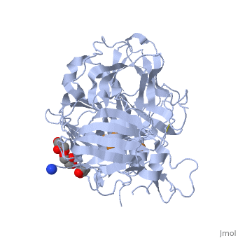
<StructureSection load='1w6l' size='340' side='right'caption='[[1w6l]], [[Resolution|resolution]] 2.00Å' scene=''> | <StructureSection load='1w6l' size='340' side='right'caption='[[1w6l]], [[Resolution|resolution]] 2.00Å' scene=''> | ||
== Structural highlights == | == Structural highlights == | ||
| - | <table><tr><td colspan='2'>[[1w6l]] is a 1 chain structure with sequence from [ | + | <table><tr><td colspan='2'>[[1w6l]] is a 1 chain structure with sequence from [https://en.wikipedia.org/wiki/"vibrio_subtilis"_ehrenberg_1835 "vibrio subtilis" ehrenberg 1835]. Full crystallographic information is available from [http://oca.weizmann.ac.il/oca-bin/ocashort?id=1W6L OCA]. For a <b>guided tour on the structure components</b> use [https://proteopedia.org/fgij/fg.htm?mol=1W6L FirstGlance]. <br> |
| - | </td></tr><tr id='ligand'><td class="sblockLbl"><b>[[Ligand|Ligands:]]</b></td><td class="sblockDat"><scene name='pdbligand=CU:COPPER+(II)+ION'>CU</scene>, <scene name='pdbligand=GOL:GLYCEROL'>GOL</scene>, <scene name='pdbligand=OXY:OXYGEN+MOLECULE'>OXY</scene></td></tr> | + | </td></tr><tr id='ligand'><td class="sblockLbl"><b>[[Ligand|Ligands:]]</b></td><td class="sblockDat" id="ligandDat"><scene name='pdbligand=CU:COPPER+(II)+ION'>CU</scene>, <scene name='pdbligand=GOL:GLYCEROL'>GOL</scene>, <scene name='pdbligand=OXY:OXYGEN+MOLECULE'>OXY</scene></td></tr> |
| - | <tr id='related'><td class="sblockLbl"><b>[[Related_structure|Related:]]</b></td><td class="sblockDat">[[1gsk|1gsk]], [[1hkp|1hkp]], [[1hkz|1hkz]], [[1hl0|1hl0]], [[1hl1|1hl1]], [[1of0|1of0]], [[1ogr|1ogr]], [[1uvw|1uvw]], [[1w6w|1w6w]]</td></tr> | + | <tr id='related'><td class="sblockLbl"><b>[[Related_structure|Related:]]</b></td><td class="sblockDat"><div style='overflow: auto; max-height: 3em;'>[[1gsk|1gsk]], [[1hkp|1hkp]], [[1hkz|1hkz]], [[1hl0|1hl0]], [[1hl1|1hl1]], [[1of0|1of0]], [[1ogr|1ogr]], [[1uvw|1uvw]], [[1w6w|1w6w]]</div></td></tr> |
| - | <tr id='resources'><td class="sblockLbl"><b>Resources:</b></td><td class="sblockDat"><span class='plainlinks'>[ | + | <tr id='resources'><td class="sblockLbl"><b>Resources:</b></td><td class="sblockDat"><span class='plainlinks'>[https://proteopedia.org/fgij/fg.htm?mol=1w6l FirstGlance], [http://oca.weizmann.ac.il/oca-bin/ocaids?id=1w6l OCA], [https://pdbe.org/1w6l PDBe], [https://www.rcsb.org/pdb/explore.do?structureId=1w6l RCSB], [https://www.ebi.ac.uk/pdbsum/1w6l PDBsum], [https://prosat.h-its.org/prosat/prosatexe?pdbcode=1w6l ProSAT]</span></td></tr> |
</table> | </table> | ||
== Function == | == Function == | ||
| - | [[ | + | [[https://www.uniprot.org/uniprot/COTA_BACSU COTA_BACSU]] Involved in brown pigmentation during sporogenesis. |
== Evolutionary Conservation == | == Evolutionary Conservation == | ||
[[Image:Consurf_key_small.gif|200px|right]] | [[Image:Consurf_key_small.gif|200px|right]] | ||
Revision as of 13:33, 13 October 2021
3D structure of CotA incubated with CuCl2
| |||||||||||