We apologize for Proteopedia being slow to respond. For the past two years, a new implementation of Proteopedia has been being built. Soon, it will replace this 18-year old system. All existing content will be moved to the new system at a date that will be announced here.
Fel d 1
From Proteopedia
(Difference between revisions)
| Line 12: | Line 12: | ||
Chains 1 and 2 are two antiparallel polypeptides, linked via 3 interchain disulfide bonds, formed between cysteine residues at positions Cys3-Cys73, Cys44-Cys48, and Cys70-Cys7, at chains 1 and 2, respectively, in chains 1 and 2, respectively<ref name="[1]"/><ref name="[4]"/>. | Chains 1 and 2 are two antiparallel polypeptides, linked via 3 interchain disulfide bonds, formed between cysteine residues at positions Cys3-Cys73, Cys44-Cys48, and Cys70-Cys7, at chains 1 and 2, respectively, in chains 1 and 2, respectively<ref name="[1]"/><ref name="[4]"/>. | ||
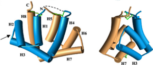
| - | [[Image:Monomero.png | thumb | 300px | '''Figure 1.'''General structure of a Fel d 1 monomer, shown in two distinct orientations, rotated 90° about the vertical axis<ref name="[1]"/>.]] | + | [[Image:Monomero.png | thumb | 300px | '''Figure 1.''' General structure of a Fel d 1 monomer, shown in two distinct orientations, rotated 90° about the vertical axis<ref name="[1]"/>.]] |
Chain 1 has about 8kDa, composed of a residue of 70 amino acids and chain 2 has about 10 kDa, which can be composed of a residue of 90 amino acids, found preferably in the sebaceous glands, or composed of a residue of 92 amino acids, which is expressed by the salivary glands. Its glycan portion is found in chain 2 and the recombinant structure of Fel d 1 reveals that the N33 residue is located in the loop connecting the H2 and H3 helices and that the side chain is exposed to the solvent<ref name="[1]"/><ref name="[4]"/>. | Chain 1 has about 8kDa, composed of a residue of 70 amino acids and chain 2 has about 10 kDa, which can be composed of a residue of 90 amino acids, found preferably in the sebaceous glands, or composed of a residue of 92 amino acids, which is expressed by the salivary glands. Its glycan portion is found in chain 2 and the recombinant structure of Fel d 1 reveals that the N33 residue is located in the loop connecting the H2 and H3 helices and that the side chain is exposed to the solvent<ref name="[1]"/><ref name="[4]"/>. | ||
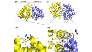
| - | [[Image:Ca2.png | thumb | 300px | '''Figure 2.'''General structure of the Fel d 1 tetramer. (a) Schematic view of the Fel d 1 tetramer (1 + 2). The two heterodimeric subunits A and B, each composed of the linked 1 and 2 chains, that form the tetramer are yellow and blue, respectively. The three Ca<sup>2+</sup> ions are indicated as red balls. (b) Schematic view of the Fel d 1 tetramer following an approximately 90° rotation about the horizontal axis. (c) Calcium binding sites 1 and 2. (d) Calcium binding site 3<ref name="[2]"/>.]] | + | [[Image:Ca2.png | thumb | 300px | '''Figure 2.''' General structure of the Fel d 1 tetramer. (a) Schematic view of the Fel d 1 tetramer (1 + 2). The two heterodimeric subunits A and B, each composed of the linked 1 and 2 chains, that form the tetramer are yellow and blue, respectively. The three Ca<sup>2+</sup> ions are indicated as red balls. (b) Schematic view of the Fel d 1 tetramer following an approximately 90° rotation about the horizontal axis. (c) Calcium binding sites 1 and 2. (d) Calcium binding site 3<ref name="[2]"/>.]] |
Figure 1 shows the general structure of a Fel d 1 monomer, shown in two different orientations, rotated 90° around the vertical axis. Chains 1 and 2 correspond to the gold and blue helices respectively. The dotted line indicates the disordered loop (residues 75 to 92). The three disulfide bridges connecting chains 1 and 2 are shown in green. An arrow indicates the unique glycosylation site at residue N33<ref name="[1]"/>. | Figure 1 shows the general structure of a Fel d 1 monomer, shown in two different orientations, rotated 90° around the vertical axis. Chains 1 and 2 correspond to the gold and blue helices respectively. The dotted line indicates the disordered loop (residues 75 to 92). The three disulfide bridges connecting chains 1 and 2 are shown in green. An arrow indicates the unique glycosylation site at residue N33<ref name="[1]"/>. | ||
| Line 24: | Line 24: | ||
=='''Treatment'''== | =='''Treatment'''== | ||
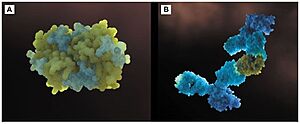
| - | [[Image:Anticorpos.jpg | thumb | 300px | '''Figure 4'''(a) Three-dimensional configuration of Fel d 1 (b) Fel d 1 linked to two anti-Fel d 1 IgY antibodies <ref name="[3]"/>.]] | + | [[Image:Anticorpos.jpg | thumb | 300px | '''Figure 4.''' (a) Three-dimensional configuration of Fel d 1 (b) Fel d 1 linked to two anti-Fel d 1 IgY antibodies <ref name="[3]"/>.]] |
Immunotherapy, or allergy vaccination, which is based on repeated subcutaneous injections of cat hair extracts has been shown to be effective in the curative treatment of allergy. However, in addition to being a time-consuming treatment, it can cause serious side effects such as asthma attacks and anaphylactic shock <ref name="[1]"/><ref name="[4]"/>. | Immunotherapy, or allergy vaccination, which is based on repeated subcutaneous injections of cat hair extracts has been shown to be effective in the curative treatment of allergy. However, in addition to being a time-consuming treatment, it can cause serious side effects such as asthma attacks and anaphylactic shock <ref name="[1]"/><ref name="[4]"/>. | ||
Additionally, to add to the immunotherapy treatment, there is a cat food supplemented with anti-Fel d 1 IgY, which significantly reduces active Fel d 1 levels. Figure 4 shows the three-dimensional configuration of Fel d 1 (a) and the structure of Fel d 1 linked to two anti-Fel d 1 IgY antibodies (b) <ref name="[3]"/>. | Additionally, to add to the immunotherapy treatment, there is a cat food supplemented with anti-Fel d 1 IgY, which significantly reduces active Fel d 1 levels. Figure 4 shows the three-dimensional configuration of Fel d 1 (a) and the structure of Fel d 1 linked to two anti-Fel d 1 IgY antibodies (b) <ref name="[3]"/>. | ||
Revision as of 13:13, 6 December 2021
1PUO - Fel d 1: The major cat allergen
| |||||||||||


