Sandbox Reserved 1716
From Proteopedia
(Difference between revisions)
| Line 1: | Line 1: | ||
| - | + | =Vitamin K Epoxide Reductase= | |
| - | + | ||
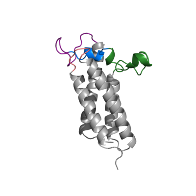
| - | <StructureSection load=' | + | <StructureSection load='6WV9' size='350' frame='true' side='right' caption='Vitamin K Epoxide Reductase 6WV9' scene=' '> |
| - | This is a default text for your page ''''''. Click above on '''edit this page''' to modify. Be careful with the < and > signs. | + | This is a default text for your page '''Kiana Jackson/Sandbox 1'''. Click above on '''edit this page''' to modify. Be careful with the < and > signs. |
You may include any references to papers as in: the use of JSmol in Proteopedia <ref>DOI 10.1002/ijch.201300024</ref> or to the article describing Jmol <ref>PMID:21638687</ref> to the rescue. | You may include any references to papers as in: the use of JSmol in Proteopedia <ref>DOI 10.1002/ijch.201300024</ref> or to the article describing Jmol <ref>PMID:21638687</ref> to the rescue. | ||
| - | == | + | == Introduction == |
| + | [[Image:VKORimage3.png|400px|right|thumb|Figure 1. Closed Conformation of VKOR due to Warfarin Binding]] | ||
| + | ===Vitamin K Cycle=== | ||
| + | ===Location of Enzyme === | ||
| + | == Structure == | ||
| + | [https://en.wikipedia.org/wiki/Vitamin_K_epoxide_reductase VKOR WIKI] | ||
| + | This is a sample scene created with SAT to <scene name="/12/3456/Sample/1">color</scene> by Group, and another to make <scene name="/12/3456/Sample/2">a transparent representation</scene> of the protein. You can make your own scenes on SAT starting from scratch or loading and editing one of these sample scenes. <ref name="Ransey">PMID:28504306</ref> | ||
| + | ===Transmembrane Helices=== | ||
| + | ===Cap Domain=== | ||
| - | == | + | ==Significant Cysteines== |
| + | <scene name='90/905622/Cysteines_43_51_residues/1'>Cysteine 43 and 51 and Residues in Between</scene> | ||
| - | == | + | ==Vitamin K Epoxide== |
| - | + | ==Warfarin== | |
| - | == | + | |
| - | + | ||
| - | + | ||
</StructureSection> | </StructureSection> | ||
== References == | == References == | ||
| + | |||
<references/> | <references/> | ||
Revision as of 19:05, 15 March 2022
Vitamin K Epoxide Reductase
| |||||||||||
References
- ↑ Hanson, R. M., Prilusky, J., Renjian, Z., Nakane, T. and Sussman, J. L. (2013), JSmol and the Next-Generation Web-Based Representation of 3D Molecular Structure as Applied to Proteopedia. Isr. J. Chem., 53:207-216. doi:http://dx.doi.org/10.1002/ijch.201300024
- ↑ Herraez A. Biomolecules in the computer: Jmol to the rescue. Biochem Mol Biol Educ. 2006 Jul;34(4):255-61. doi: 10.1002/bmb.2006.494034042644. PMID:21638687 doi:10.1002/bmb.2006.494034042644
- ↑ Ransey E, Paredes E, Dey SK, Das SR, Heroux A, Macbeth MR. Crystal structure of the Entamoeba histolytica RNA lariat debranching enzyme EhDbr1 reveals a catalytic Zn(2+) /Mn(2+) heterobinucleation. FEBS Lett. 2017 Jul;591(13):2003-2010. doi: 10.1002/1873-3468.12677. Epub 2017, Jun 14. PMID:28504306 doi:http://dx.doi.org/10.1002/1873-3468.12677

